The counting rule
A mitosis animation should show DNA replication before mitosis, then the separation of sister chromatids during anaphase. A replicated chromosome has two sister chromatids. Once the sisters separate, each is counted as a chromosome. Always say whether a number describes the whole dividing cell, one spindle pole or one daughter cell.
For teachers, the difficult moment is often a pause between metaphase and anaphase. The DNA has not suddenly doubled again, yet the chromosome count across the still-undivided cell changes. A useful animation makes that change in counting visible without making new DNA appear.
This guide uses a deliberately simplified animal-cell example with four chromosomes before replication, written as 2n = 4. It is a storyboard you can develop in Animiotics, with a count ledger and classroom questions. The CGI images are conceptual illustrations, not microscope observations or measured chromosome structures.
Name the objects before moving them
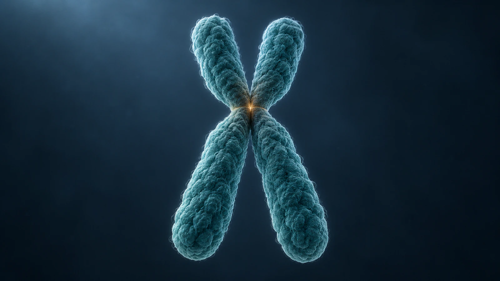
A chromosome is a DNA-containing structure; a chromatid is one of the two copies in a replicated chromosome. The NHGRI chromatid definition describes the sisters as joined until they separate during division. Do not use “chromosome” as a synonym for every visible arm of an X.
In a condensed replicated chromosome, each sister extends along one side of the model and has two arms. The central constriction corresponds to the centromeric region. The NHGRI centromere glossary helps distinguish this region from the chromosome arms; it is not a cut that divides a chromosome into an upper and a lower half.
Give the scene objects working names before adding motion: chromosome A, sister A1 and sister A2, for example. Keep those names in a separate production note even if the audience never sees them. This makes an accidental duplicate or missing sister easier to identify during review.
Choose one counting boundary
Use the entire cell as the boundary through metaphase. At anaphase, report both the total in the undivided cell and the number moving toward each pole. After cytokinesis, the physical division of the cell, return to a per-cell count. Changing the boundary without saying so is a common way to make a correct diagram feel contradictory.
For this example, 2n = 4 means two homologous pairs. Homologs are the corresponding maternal and paternal chromosomes, whereas sisters are replicated copies of one chromosome. Use a long pair and a short pair to make the two homologous pairs easy to recognize. Do not make one whole chromosome disappear behind another during a counting pause.
The table is a bookkeeping model for normal segregation, not a statement that all animal cells have four chromosomes. DNA molecules here means chromosomal double-stranded DNA molecules; the count excludes mitochondrial DNA. In the final row, each daughter cell is considered separately.
| Pause point | Counting boundary | Chromosomes | Chromosomal DNA molecules |
|---|---|---|---|
| Before S phase | One cell | 4 | 4 |
| After S phase / metaphase | One cell | 4, each with two sisters | 8 |
| Anaphase | Whole undivided cell | 8 separated chromosomes | 8 |
| Anaphase | One pole’s destination set | 4 | 4 |
| After division | Each daughter cell | 4 | 4 |
Keep replication outside mitosis
OpenStax Biology 2e places DNA replication in S phase, before mitosis. Mitosis distributes the replicated chromosomes into daughter nuclei. An introductory movie can begin at metaphase, but it should then state that replication has already occurred.
If you include an earlier scene, distinguish an explanatory model from the appearance of interphase chromatin. A row of compact X shapes is convenient for counting but should not imply that chromosomes remain that visibly condensed throughout the cell cycle. A brief transition can establish the counting relationship without claiming to depict the molecular replication machinery.
In the production note, mark the replication transition as an illustrative summary. Avoid a dissolve that creates extra chromosomes during anaphase. If the purpose is chromosome counting rather than replication chemistry, a short statement about the completed S phase is clearer than an unrelated close-up of a DNA helix.
Design the separation correctly
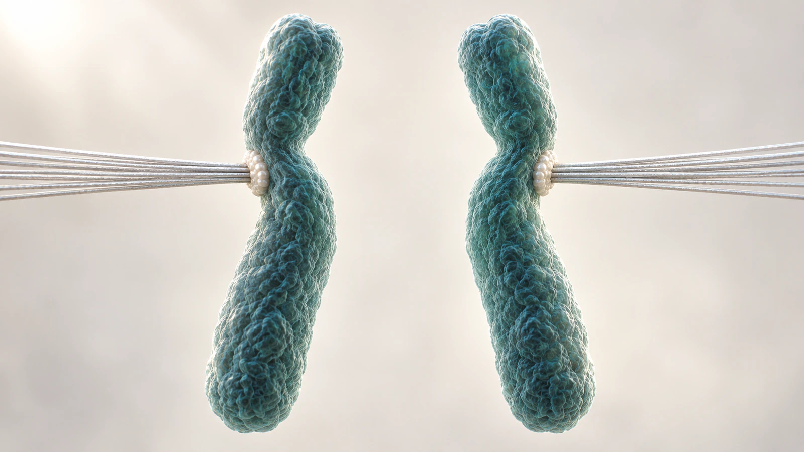
The critical visual event is two sister bodies moving apart, not an X being cut across its middle. Each departing body must preserve both arms of the sister it came from. At separation, change the narration to “daughter chromosomes” while preserving the same visible objects.
OpenStax describes spindle attachment through kinetochores, protein structures at the centromeric regions, and the movement of separated sisters toward opposite poles. For a simple classroom shot, show a restrained set of spindle fibers and clear attachment regions. A filament that passes through a chromosome arm can teach the wrong connection even when the overall scene looks attractive.
Keep the camera fixed for the first separation pass. A dramatic orbit can hide an object just as the viewer tries to count it. Once the count has been established, a second view can reveal spatial depth. These are presentation choices, not measurements of spindle speed or a claim that all chromosomes move synchronously.
Build a six-shot lesson
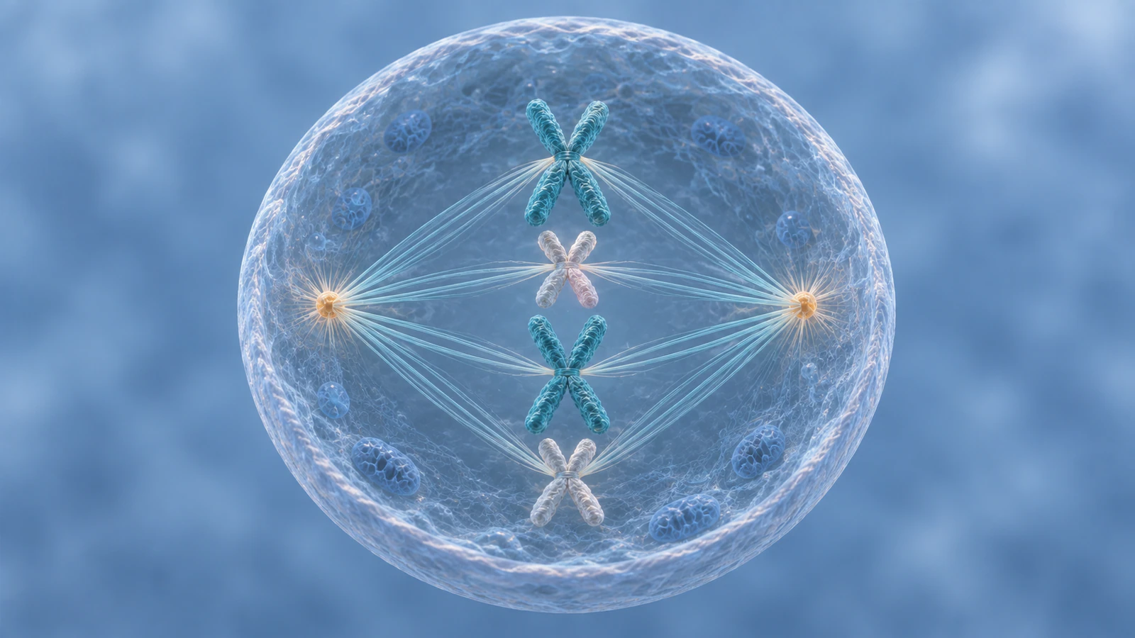
Use the following sequence as an authoring plan, with holds long enough for your learners to answer. Do not treat the suggested scene order as a measured time course. The original contribution is the explicit boundary and object audit at each pause, which can be reused with another chromosome number.
Create a simple review sheet with a row for each shot. Record the visible objects, the narration’s count and the reason for any change. A mismatch is a concrete correction request: “Keep four replicated chromosomes in this view,” rather than “Make the biology more accurate.”
| Shot | What viewers see | Question to ask |
|---|---|---|
| 1. Establish | Four schematic unreplicated chromosome identities | What does our 2n = 4 model represent? |
| 2. Replication summary | Each identity now has two sister copies | Did the chromosome count or the DNA amount change? |
| 3. Metaphase hold | Four replicated chromosomes with readable sisters | Can you find eight chromatids? |
| 4. Anaphase start | The same sisters separate without new objects | What are the separated sisters called now? |
| 5. Two destination sets | Four daughter chromosomes toward each pole | Are you counting one pole or the whole cell? |
| 6. Division and reset | Two daughter cells, then a simplified count recap | What does each cell inherit? |
Use Animiotics with a count ledger
Animiotics is a product for creating scientific animations with AI. Its current documentation also describes an editable “Mitosis & daughter cells” starter with six replicated chromosomes. That starter and this four-chromosome exercise use different counts. Either keep the starter’s six consistently or build the four-chromosome exercise; do not silently switch between them.
When using the starter library, preview the example and create your own editable copy through the documented customization flow. Inspect the chromosome objects and the full timeline before adapting the lesson. A finished preview and the real-time editor view can differ in materials, so check your own result rather than judging only the library thumbnail.
For an AI-created scene, make the count and object continuity explicit in the prompt. Review the generated result against the ledger before accepting its appearance. The example below is a proposed brief, not a tested prompt or a guarantee that one generation will preserve every chromosome. Account access and the usage terms shown in the product apply.
A prompt with a reviewable target
Proposed brief: “Create a conceptual animal-cell mitosis explanation for a biology class. Start after replication with four replicated chromosomes, each made of two sister chromatids. Use two long and two short chromosome identities. Hold at metaphase with every chromosome visible. Separate the existing sisters toward opposite poles so that four daughter chromosomes reach each pole. Do not create new DNA during separation. Keep the camera steady for the counting holds. Use restrained colors and a clear cell boundary. Geometry and timing are illustrative.”
Treat that brief as a specification to check, not evidence that the output is correct. Scrub the first separation frames and follow one sister from its original position to its destination. Repeat for the others. If a sister changes identity, vanishes or becomes a new X, correct that transition before adding camera movement.
For a broader scene-planning method, use the biology animation storyboard guide. For constraints and revision language, see the scientific animation prompt guide. Here, the acceptance criterion is especially concrete: the same eight chromosomal DNA copies must be accounted for across the separation.
Color identity, not the answer
Choose colors that help students track an identity rather than guess the phase. Keep both sisters of one chromosome visually related and preserve their appearance after separation. Distinguish homologs with a second cue such as a subtle surface pattern or a consistent spoken name, so the lesson does not rely on color alone.
A teal object should not turn amber merely because it has crossed the cell center. That would introduce a change students may interpret as a biological transformation. Likewise, brightness should not stand for DNA amount unless you have explicitly defined an illustrative convention.
Review the lesson at the actual classroom display size. If the short chromosome is hidden by a caption or a bright spindle bundle, the count is not usable. Favor clear spacing over dense cytoplasmic detail during the pause frames. You can restore surrounding context in the transition between those holds.
Test the explanation with three pauses
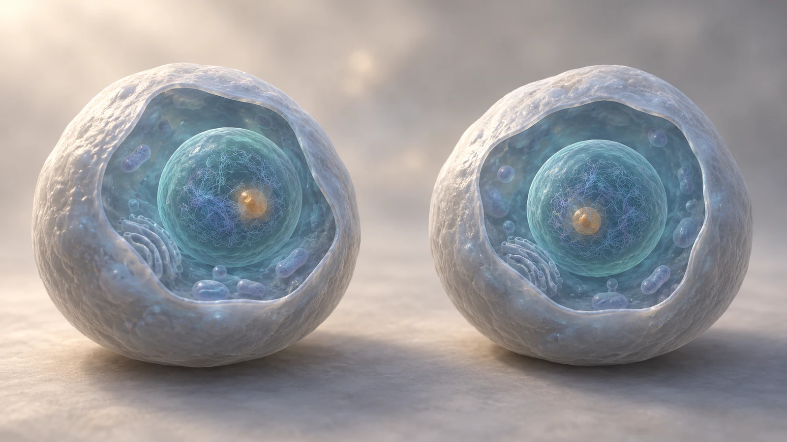
At metaphase, ask learners to write two numbers and their units: chromosomes per cell and chromatids per cell. At anaphase, ask for chromosomes in the whole cell and chromosomes heading to one pole. After division, ask for chromosomes per daughter cell. Keep the boundary visible or state it aloud every time.
Then ask one transfer question: “If the starting cell had six chromosomes instead of four, which numbers would change?” A learner who can apply the rule to the documented six-chromosome starter has understood more than the particular colors in your movie. Do not present this exercise as a validated assessment instrument or promise a learning gain.
Finish the visual audit at cytokinesis. The daughter nuclei should receive corresponding sets without an extra replication event. If you show chromosomes decondensing, use a separate recap to count their identities rather than pretending that distinct X shapes remain visible in the new interphase nuclei. Preserve the distinction between a teaching overlay and the biological view.
Frequently asked questions
Is an X-shaped chromosome one chromosome or two?
Before sister separation, it represents one replicated chromosome made of two sister chromatids. The four visible arms are not four chromosomes.
Does chromosome number double during DNA replication?
In the usual counting convention, replication doubles the chromosomal DNA molecules and produces sister chromatids while the chromosome count remains unchanged until the sisters separate.
Why does anaphase have eight chromosomes in the four-chromosome example?
The separated sisters are now counted individually, so the still-undivided cell contains eight daughter chromosomes, with four moving toward each pole. No additional DNA replication has occurred.
Should the animation show the same count as the Animiotics starter?
It should use one declared count consistently. The documented starter uses six replicated chromosomes, while this hypothetical teaching plan uses four; adapt the ledger to whichever model you actually use.
Can an AI-generated mitosis animation be used without scientific review?
Review the counts, sister continuity, attachment geometry and final destination sets first. A convincing render does not establish biological accuracy, and a prompt is not proof that every frame meets it.
Try your own counting lesson
Start with one metaphase hold and one separation shot in Animiotics. Write the expected counts before creating the scene, then trace every sister through the transition. Once that small sequence is correct, expand it into a classroom explanation with a question at each pause.