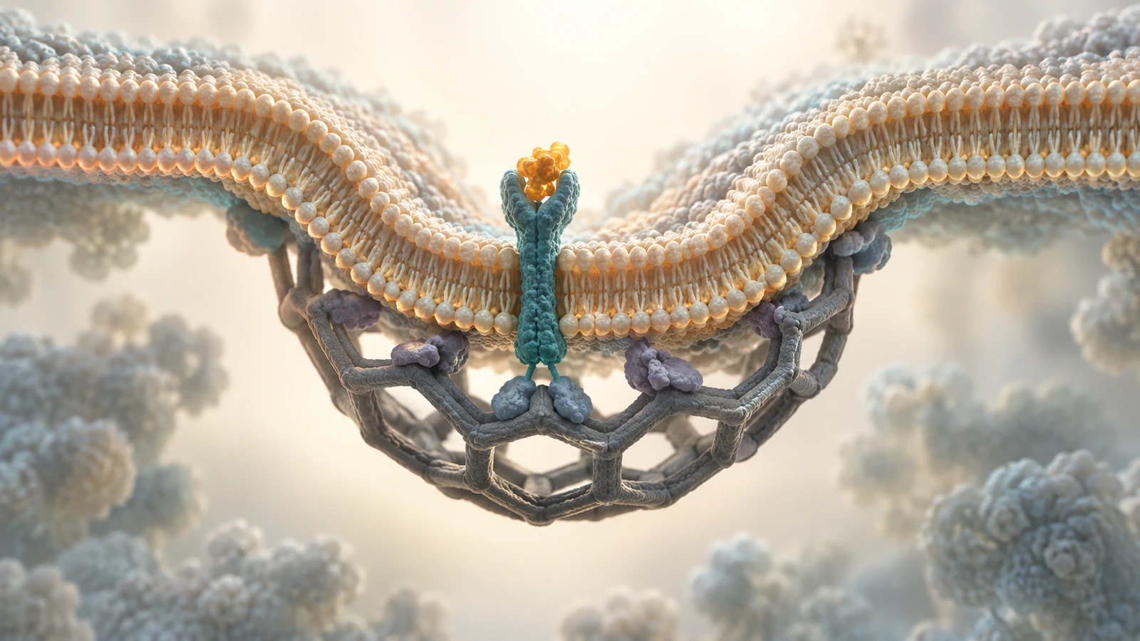
Begin with the student question
A biology animation storyboard is a sequence of planned visual states, not a list of attractive shots. Start by writing the question students should answer after watching. For the example in this guide, the question is: how can a cell bring a selected surface-bound cargo inside without turning the plasma membrane into an open hole? The answer requires a membrane pocket, a still-connected neck and then a sealed vesicle.
The storyboard records what a viewer must see at each step, what source supports it and what the image does not claim. A teacher can use it before generating a scene in Animiotics and again while reviewing the draft. The same method works for a research presentation, but the vocabulary and pacing here are aimed at a classroom.
The clathrin-mediated uptake shown below is a conceptual example, not a reconstruction of a particular receptor, measured cell or experiment. Colors and shot durations are teaching choices. AI can propose geometry and motion; a human still has to check them against the biology.
Separate evidence from illustration
Find the smallest defensible claim before drawing a frame. In clathrin-mediated endocytosis, selected cargo can be taken from the cell surface into a clathrin-coated vesicle. The cargo may be a membrane receptor and a ligand bound to its extracellular side. Adaptor proteins help connect membrane-associated cargo to clathrin on the cytosolic face, according to the Journal of Cell Biology review.
That is enough to plan a useful visual relationship. It is not enough to say that ligand binding always starts the pit or that one adaptor works identically in every organism. The review describes uncertainty in how sites initiate and differences between systems. Put such qualifications in the scene record, even if the final classroom narration is simpler.
Give each claim one visible consequence. For example, if the claim is that clathrin is on the cytosolic side, its coat must never jump to the extracellular surface between shots. If the claim is that cargo is internalized in a vesicle, the same ligand and receptor should remain trackable as the membrane bends. This converts scientific review into something a viewer can actually inspect.
Choose the teaching model
A source may describe many participants and alternative pathways. Choose one model and state its boundaries: a generic clathrin-mediated surface uptake sequence in a eukaryotic cell. Avoid naming a receptor or presenting exact molecular shapes without appropriate structural evidence.
RCSB PDB-101 describes clathrin as a coat made from three-legged components and explains that the coat disassembles after vesicle release. It also explains the role of adaptors between clathrin and membrane. That supports a sparse coat as a visual cue, not an arbitrary cage covering both sides of the bilayer. The RCSB clathrin overview is a useful reference for the relationship; it does not make the conceptual artwork on this page coordinate-derived.
Set the same orientation across the sequence. Place extracellular fluid above the plasma membrane and cytosol below it in the opening view. The receptor spans the bilayer, its ligand-binding part faces outward, and its cytosolic tail faces the adaptor and coat. Camera motion can change later, but the storyboard must explicitly preserve those relationships.
Make a source-to-scene record
Before moving anything, write one row per claim or transition. The columns below are a compact review contract. They prevent a familiar failure: a shot looks plausible on its own but contradicts the preceding shot or silently upgrades a teaching simplification into a measured mechanism.
For a teacher, the final column can also become a discussion prompt. Ask students which feature would falsify the drawing: a coat outside the cell, cargo vanishing at the neck or an open vesicle after release. Their answers test whether the motion taught the intended relationship rather than merely looked polished.
| Claim or beat | Source basis | Required visible state | Motion or camera | Check and limit |
|---|---|---|---|---|
| Cargo is selected | Receptor and adaptor accounts | Same ligand remains with one receptor | Hold a readable exterior view | Do not imply all ligands use this pathway |
| Coat supports a pit | RCSB and JCB review | Sparse coat on cytosolic face | Move to a side cutaway | Adaptor link is a simplification |
| Pit has not released | Endocytosis stage review | Neck remains continuous | Pause before scission | No universal dwell time implied |
| Vesicle is released | Stage and uncoating review | Closed bilayer around cargo | Isolate one vesicle | Do not show an automatic endosomal fate |
Beat one: establish inside and outside
Open with enough membrane to show both sides at once. A narrow crop of a receptor alone will not tell students whether the amber cargo sits outside the cell or inside it. Keep the receptor in the membrane and hold the exterior ligand long enough for the viewer to connect the two objects. Only then move to a closer view of the adaptor.
The first frame should answer a small question: what crosses the membrane and what stays on one side? Use color for identity, not for unsupported chemistry. A muted teal receptor can retain its color through all beats; an amber ligand should not become a new amber particle in the cytosol without a visible route. This identity rule is also useful when turning a PDB structure into an animation: a camera change should not silently change the molecule.
In a classroom, pause this first beat and ask students to point to the cytosolic face before any budding occurs. If they cannot, the visual foundation is too ambiguous. Reframe the scene rather than add a decorative arrow that only works while the arrow remains on screen.
Beat two: link receptor, adaptor and coat
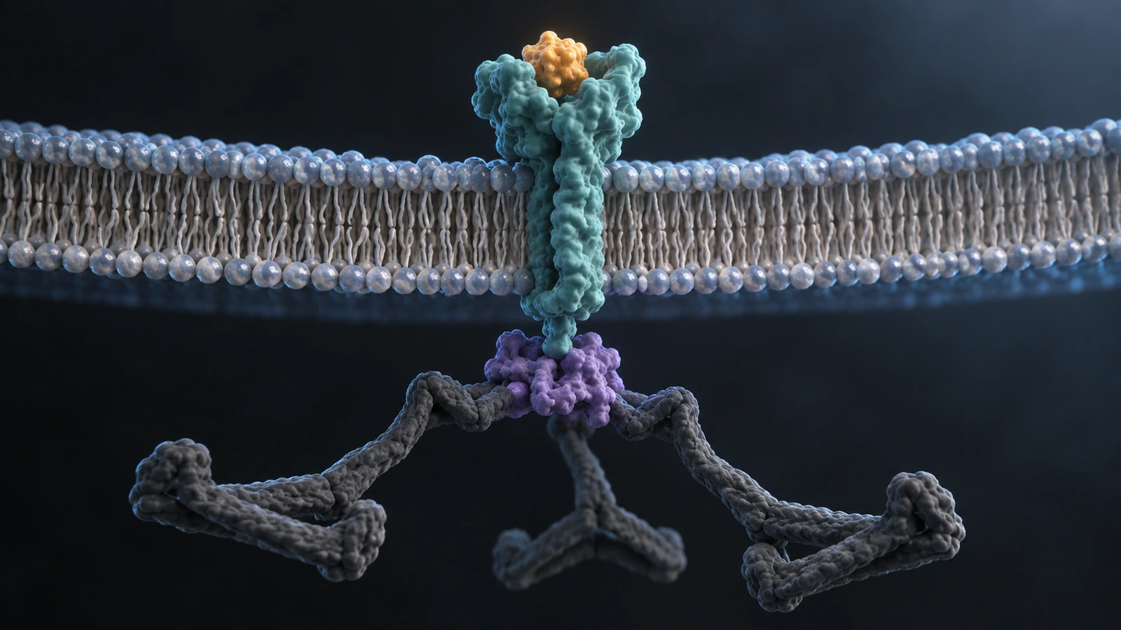
Move close enough to distinguish three jobs. The receptor spans the lipid bilayer and carries the extracellular ligand. An adaptor sits on the cytosolic side and helps connect membrane-associated cargo to clathrin. Clathrin forms a coat outside the budding pocket, on the cytosolic face. The JCB review explicitly treats adaptor, cargo and lipid contributions to early site formation.
Do not draw clathrin as a hand that directly grabs the ligand across the membrane. Also avoid a single rigid claim that adaptor recruitment always follows ligand binding; the literature does not support that universal sequence. A teaching storyboard can hold these relationships as a representative state and describe the order as simplified.
This is the moment for a restrained camera move, not a rapid orbit. Keep the membrane plane legible. If the receptor tail disappears behind a dense coat, reduce coat detail so the adaptor link remains visible. More molecular objects do not automatically make a scene more informative.
Beat three: keep the pit connected
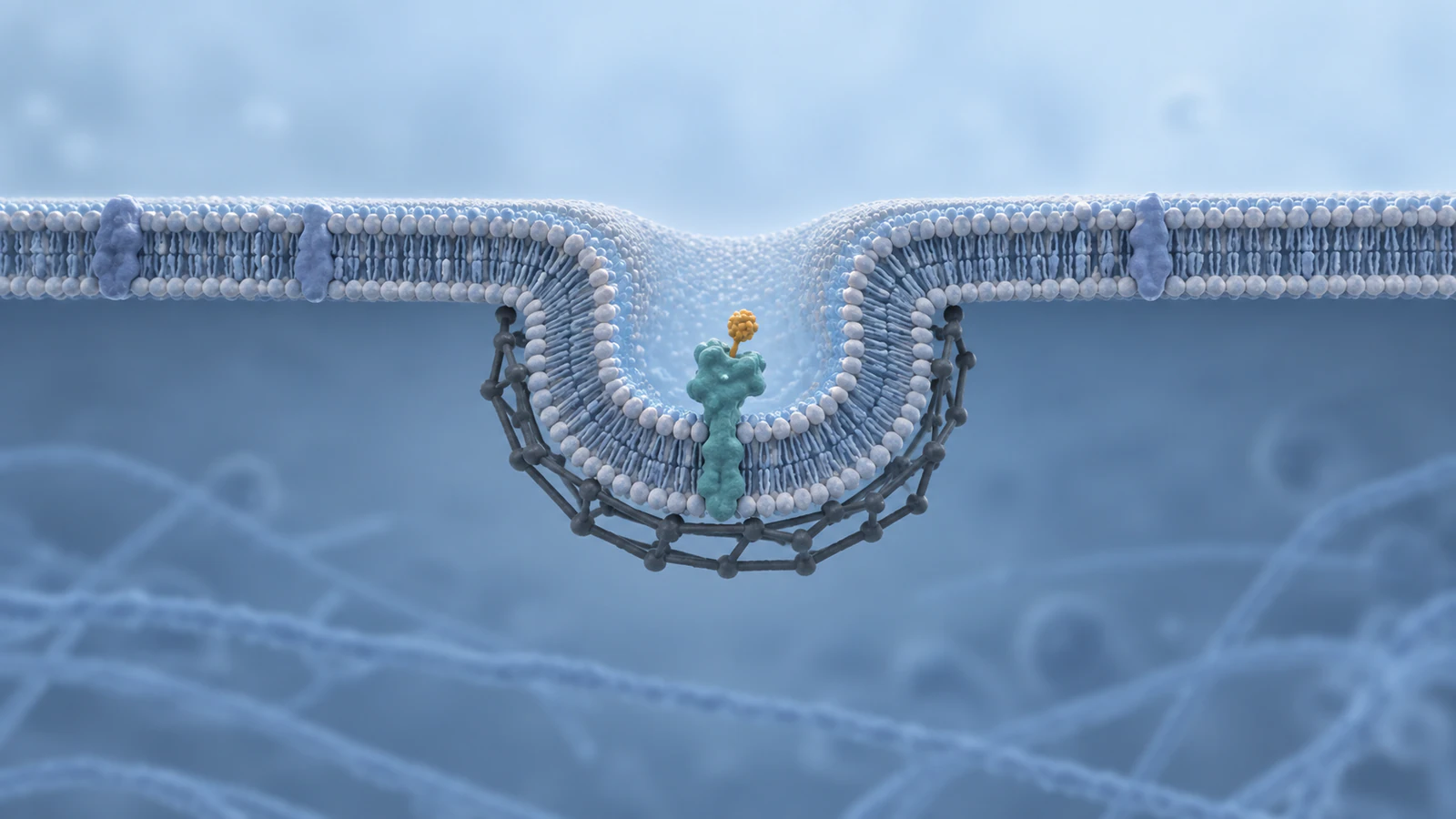
Show the membrane bending inward while it remains one continuous bilayer. Until scission, the aqueous pocket still opens to the exterior through a neck. That topological fact is more important to the lesson than a precise count of clathrin struts. A wide side cutaway makes the neck inspectable; a fully covered frontal view may hide it.
The Frontiers review separates initiation, growth, stabilization, budding, scission and uncoating as useful stages. Treat those divisions as a planning scaffold rather than a measured clock. An image of a deep pit belongs before the image of a free vesicle, even if an edit compresses the transition.
There is a known visual trap here: the lipids at the pit rim may look like two unrelated walls if the fold is drawn too thick or the camera clips the rim. Trace the membrane from left to right in every planned frame. If a student cannot follow one continuous boundary into the pocket, revise the angle or simplify the geometry.
Beat four: release, then uncoat
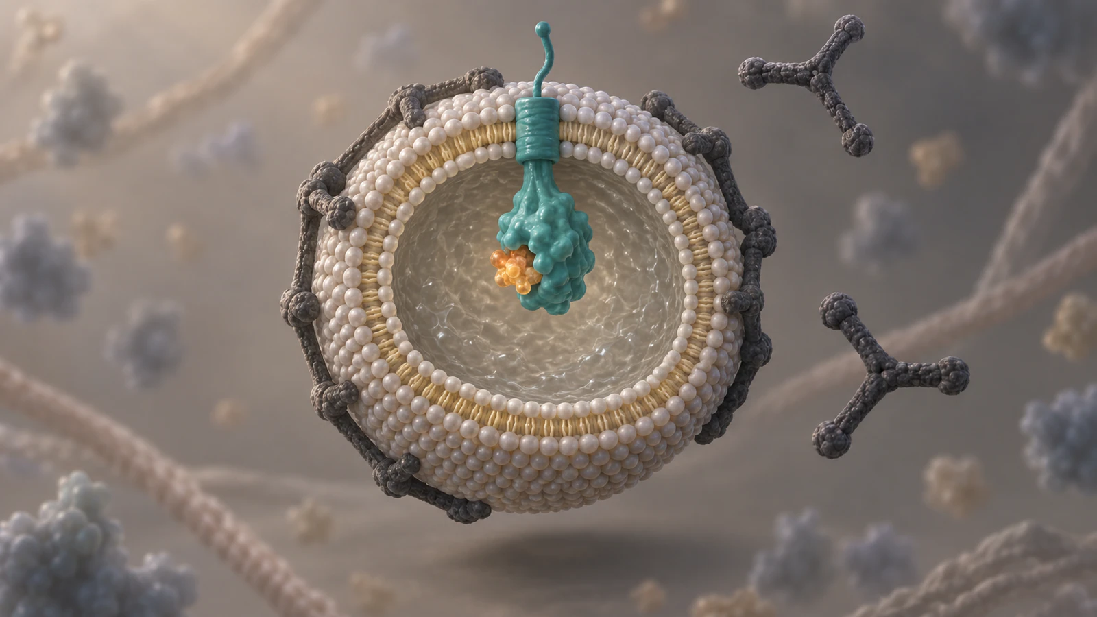
After scission, show one closed vesicle in the cytosol. The receptor still spans its membrane. Its former extracellular ligand-binding part now faces the vesicle lumen, where the ligand remains enclosed. Clathrin is on the vesicle's outer cytosolic surface and can start to come away. This is the orientation check that often exposes a beautiful but biologically reversed render.
RCSB describes coat removal after budding. The Frontiers account places uncoating after scission in its sequence. For this lesson, stop at a released, partly uncoating vesicle. Do not imply that every cargo then takes the same route to a lysosome or back to the surface.
If you decide to continue into endosomal sorting, research that as a separate scene contract. The existing endocytosis explainer gives broader communication context; this storyboard focuses on the membrane event students must see clearly.
Set camera and timing for comprehension
A storyboard should state what the camera reveals, not just where it moves. Start with a side view that establishes orientation, use a close view for the receptor-adaptor relationship, return to a wider profile for the pit neck and isolate the free vesicle for the final state. The four camera decisions correspond to four questions. Avoid rotating around the bilayer so fast that inside and outside swap in the viewer's mental model.
Write a pause into the transition before scission. That hold lets students predict what must change to produce a sealed vesicle. The visual rhythm can be slow even when real molecular events have different measured kinetics; the animation is a teaching explanation, not a time-lapse claim. Do not put a numeric duration on the screen without a source for the system being shown.
If the lesson will have narration, draft one sentence per beat before rendering. A published science-animation production account separates script, storyboard, draft review and delivery. That is a practical reminder to test the explanation while changes remain cheap, not evidence that one prescribed production order fits every class.
Draft and edit in Animiotics
Use the record as the creation brief. In Animiotics, describe one cell-surface membrane, one receptor with a bound exterior ligand, a restrained cytosolic coat and the transition from connected pit to closed vesicle. Specify the camera's side view and the color identity you want preserved. The scientific animation prompt guide has a fuller prompt worksheet if you need to separate subject, action, exclusions and review criteria.
Then inspect the generated scene beat by beat. Edit the camera, material and timing so the membrane boundary stays readable. If the receptor flips orientation or the coat appears on the wrong side, correct that state before polishing lighting. The current product lets you describe an animation and work with the scene, but this proposed classroom sequence has not been tested as an exact Animiotics output. Do not treat the first AI result as scientific validation.
Keep one cargo, one receptor and one pit in the first pass. Add another molecule only if it answers a student question. Save a still from each beat for review, and note where a cut could create a false biological transition.
Review the draft with a purpose
Ask a subject expert to check the claim ledger and a student or colleague to explain what each transition shows. Those are different reviews. The expert may detect an incorrect adaptor relationship; the student may reveal that the neck was never visible. Both observations tell you what to edit. Neither is a substitute for checking the primary evidence behind a detailed scientific claim.
Use a short pass/fail checklist: can viewers identify extracellular and cytosolic faces, follow the same ligand and receptor, see the coat on the correct side, tell when the neck remains open and tell when the vesicle closes? Also check that captions identify the images as conceptual and do not imply a measured receptor structure or universal kinetics. If a frame fails, repair that frame or the adjoining cut, not the entire visual language.
Finally, compare the export at the actual classroom screen size. Thin receptor tails and dark coats can vanish in a small projected image. A clear silhouette and a well-timed pause often teach more than another decorative particle. Keep the key question visible in the teacher's discussion, even if the video itself carries no on-screen label.
Frequently asked questions
What belongs in a biology animation storyboard?
Put the student question, source-backed claim, required visible state, camera or motion plan, and scientific caveat in each beat. For a membrane process, track inside/outside orientation and object identity across cuts.
Does a storyboard need exact molecular structures?
No. Use an exact structure when the lesson depends on its geometry and you can verify the identifier and biological assembly. A conceptual form is appropriate for teaching topology if you label it honestly and avoid pretending its fold is measured.
Does clathrin pull directly on cargo?
A clathrin coat is connected to membrane-associated cargo through adaptor and accessory proteins. A simplified scene should keep that relationship clear rather than placing clathrin on the extracellular ligand.
When should the vesicle become closed?
After the pit neck is cut in the planned sequence. Before that transition, draw a continuous opening to the cell exterior; after it, draw a sealed membrane around cargo in the cytosol.
Can an AI-made animation be used without review?
It can be a draft, but it still needs scientific and teaching review. Check source claims, topology, molecular identity, captions and what a student actually infers from the motion.
Try one classroom sequence
Start with the four-state clathrin uptake plan: surface cargo, adaptor and coat, connected pit, closed vesicle. Build a short draft in Animiotics, then pause at the neck and ask what must change next. Revise the scene until the answer is visible without a misleading arrow or unsupported detail.