Show exchange before volume
A clear osmosis animation shows water crossing a selectively permeable membrane, then connects the net movement to a change in cell volume. Individual water molecules can move in both directions. The useful question is which direction has the greater overall transfer under the conditions being illustrated, not whether every molecule follows the same arrow.
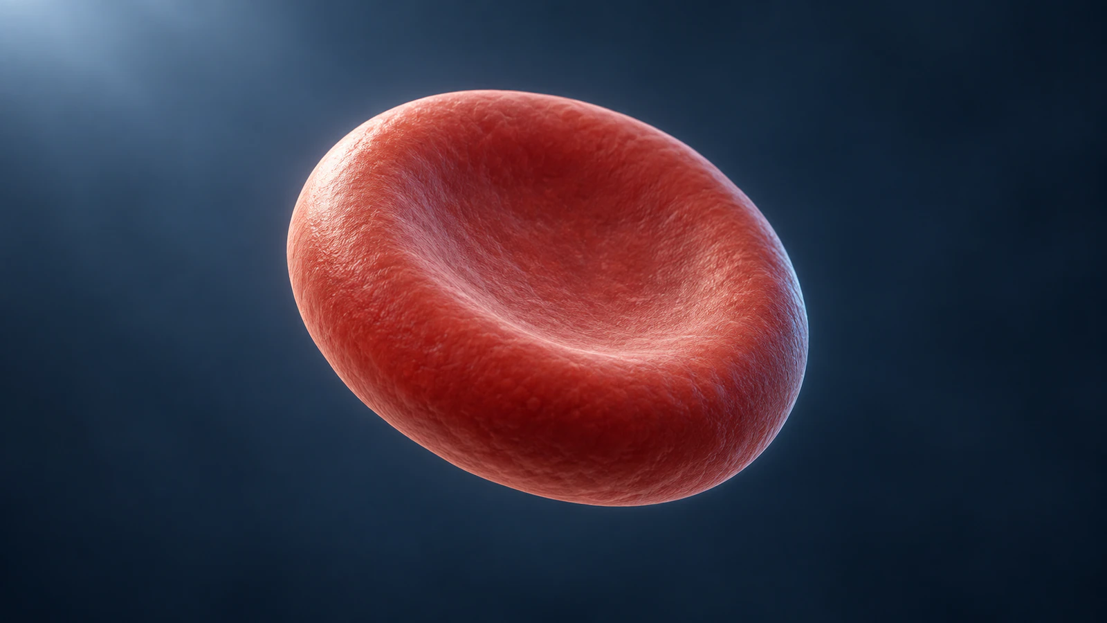
This guide is for biology students making a short class presentation or study-group explanation. You can use the scene plan in Animiotics to create an editable scientific animation, then check whether the finished sequence actually teaches the relationship you intended. The worked example uses a conceptual red blood cell and deliberately simple surrounding solutions.
Treat the molecular view and the whole-cell view as two linked explanations. One shows exchange across a boundary; the other shows a qualitative outcome. The illustrations here are independently created conceptual views, not microscopy, molecular simulations or measured stages of one experiment.
Write the membrane rule first
Before drawing particles, write what is allowed to cross the membrane. For the basic example, water can cross and the chosen solute particles cannot cross during the interval shown. A solute is a dissolved substance; a selectively permeable membrane allows some substances to pass more readily than others.
Under that simplified rule, with no opposing pressure difference imposed, net water transfer is toward the side with more nonpenetrating solute particles per volume. The OpenStax passive-transport chapter explains the membrane constraint and the role of pressure in limiting osmosis. This qualification belongs in the setup, before a viewer starts interpreting the motion.
Make the rule an instruction to the animation, not a footnote added afterward. If the solute quietly drifts through the boundary, the scene has changed its own experiment. If the membrane opens a giant hole, the viewer can no longer tell whether selective transport is the point. Review these relationships before choosing a more dramatic camera move.
Keep two tracks separate
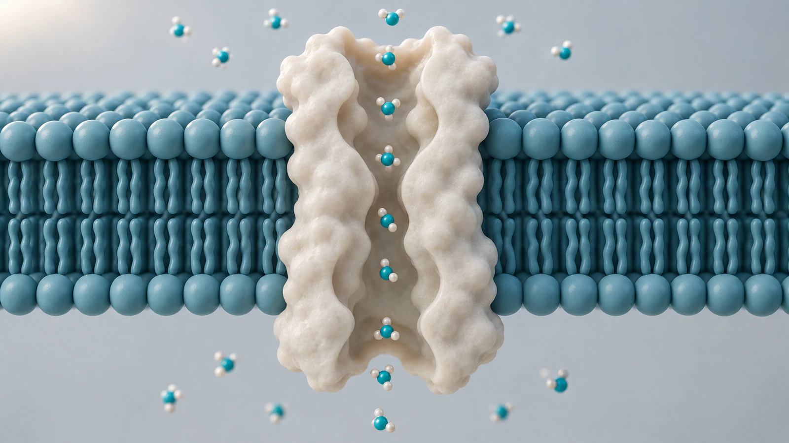
Build the sequence around two tracks: molecular exchange and cell shape. In the exchange track, a small number of representative water objects cross in both directions. In the shape track, the cell changes only enough to communicate the chosen qualitative outcome. The visible particles are teaching symbols, not an inventory of the cell's water.
Do not make each displayed particle trigger a visible bump or a fixed increment of expansion. That creates an unsupported conversion between an illustrative count and a measured volume. Instead, use narration or a brief pause to connect the overall imbalance with the later whole-cell view.
For a small presentation screen, test the exchange track without glow, trails or decorative particles. A viewer should still locate the boundary and identify which objects can cross. The diffusion and directed-transport guide offers a complementary check on motion language; this example adds a specific membrane rule and a volume consequence.
Use tonicity as a comparison
Tonicity describes how a surrounding solution affects cell volume relative to the cell and its membrane. Osmolarity counts dissolved particles per volume of solution; the two terms are not interchangeable when some solutes can cross. State the comparison and the permeability assumptions whenever you label a condition.
The 2025 review by Kuang and colleagues examines why tonicity terminology confuses learners and emphasizes membrane dependence. It also proposes changes to terminology. You do not need to adopt those proposals to improve a student animation: specifying the membrane and the nonpenetrating solutes already removes an important ambiguity.
Avoid naming the background color “hypertonic” as though blue fluid has that property by itself. Write a complete production note instead: the surrounding solution is hypertonic relative to this cell under the stated membrane conditions. That note tells a reviewer what comparison the shot is meant to communicate and which assumption would change the prediction.
Plan the comparison branches
Use the same initial cell, framing and lighting for each branch of your animated comparison. Reset to that starting state before changing the surrounding condition. A shrinking cell followed immediately by a swelling cell can otherwise look like one continuous treatment sequence, which is a different story.
The following table is a storyboard for the simplified model, not a recipe for a laboratory solution. Introduce the condition verbally or in the presentation's accessible text. Keep the biological render uncluttered so students can follow the boundary and the changing silhouette.
| Branch | What the model illustrates | What to check in the edit |
|---|---|---|
| Isotonic surroundings | No sustained net volume change, with continuing water exchange | Do not freeze all molecular movement |
| Hypotonic surroundings | Water gain and swelling; sufficiently severe challenge may cause lysis | Do not make bursting the inevitable ending of every example |
| Hypertonic surroundings | Water loss and a smaller, potentially crenated red cell | Keep the camera fixed so scale change is not a zoom |
| Reset between branches | Return to the same declared initial condition | Use a clear pause or transition, not an unexplained reversal |
Cut deliberately between scales
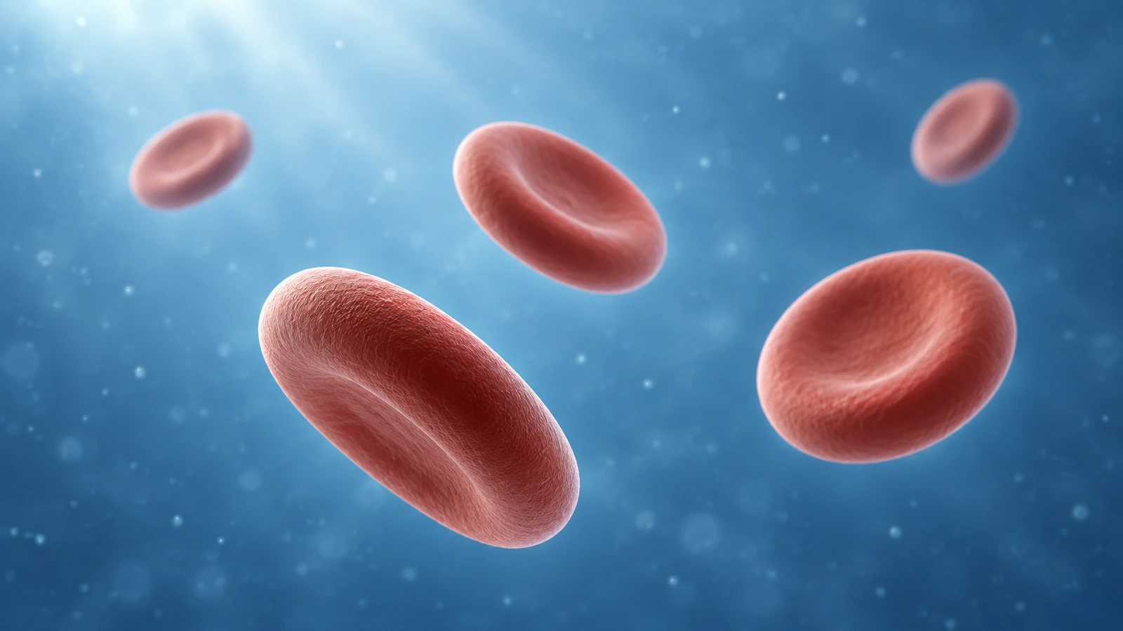
Start with a whole-cell view that makes the surrounding fluid visible as space around the cell. Then cut to a local membrane view. A deliberate change of shot is easier to interpret than a camera dive that leaves giant water molecules floating beside a complete red cell.
Use the wide context image to establish that the cell has an outside. Its few cells are a composition choice, not a sample count or a concentration estimate. When the view changes scale, say so. Do not ask the audience to infer a molecular scale from texture alone.
For the close-up, preserve the two sides of the membrane even if you remove part of it to reveal a pathway. The membrane-cutaway guide explains how to keep inside and outside legible. Keep that orientation consistent when you return to the cell, rather than rotating the boundary until the direction of exchange becomes ambiguous.
Give the water channel a role
Aquaporins are membrane proteins that provide pathways for water. A close-up can help explain how a membrane can be a barrier while still permitting water exchange. It should not turn the protein into a mechanical pump pushing a procession of molecules in one direction.
The RCSB PDB-101 aquaporin article describes the familiar four-subunit arrangement with a water pore in each subunit. Do not depict the central space between all four subunits as their single shared water pipe. The mechanism illustration here isolates one conceptual subunit to keep that teaching view simple.
For an introductory presentation, omit detailed residue labels and exact water orientations unless they are part of the question. If you later need an atomic explanation, replace the conceptual geometry with an appropriate verified structure and document its identity. More surface detail in a generated image does not make its geometry a structural measurement.
Build a six-shot explanation
Keep each shot responsible for one question. The sequence below can support a short spoken explanation without forcing molecular exchange and whole-cell shape into the same frame. Its order is a teaching proposal; the durations should follow your narration rather than pretend to reproduce biological time.
Pause after the initial condition and ask the audience to predict the next whole-cell view. That pause gives the animation a job beyond showing an attractive transformation. It also gives you a simple way to test whether the membrane rule was clear enough.
| Shot | Visual task | Question to answer |
|---|---|---|
| 1. Establish | Show the intact red cell in its surrounding medium | Which compartment is outside? |
| 2. Specify | Cut to the membrane and distinguish water from retained solute | What can cross during this example? |
| 3. Exchange | Show crossings in both directions with a declared net imbalance | What does net movement mean? |
| 4. Connect | Return to the same whole-cell camera and show the qualitative change | How does the exchange affect this model cell? |
| 5. Reset | Return to the initial state and contrast another condition | Which changed assumption reverses the prediction? |
| 6. Test | Hold an unlabeled condition while the learner predicts the outcome | Can the viewer explain the result without guessing from color? |
Check the endpoint honestly
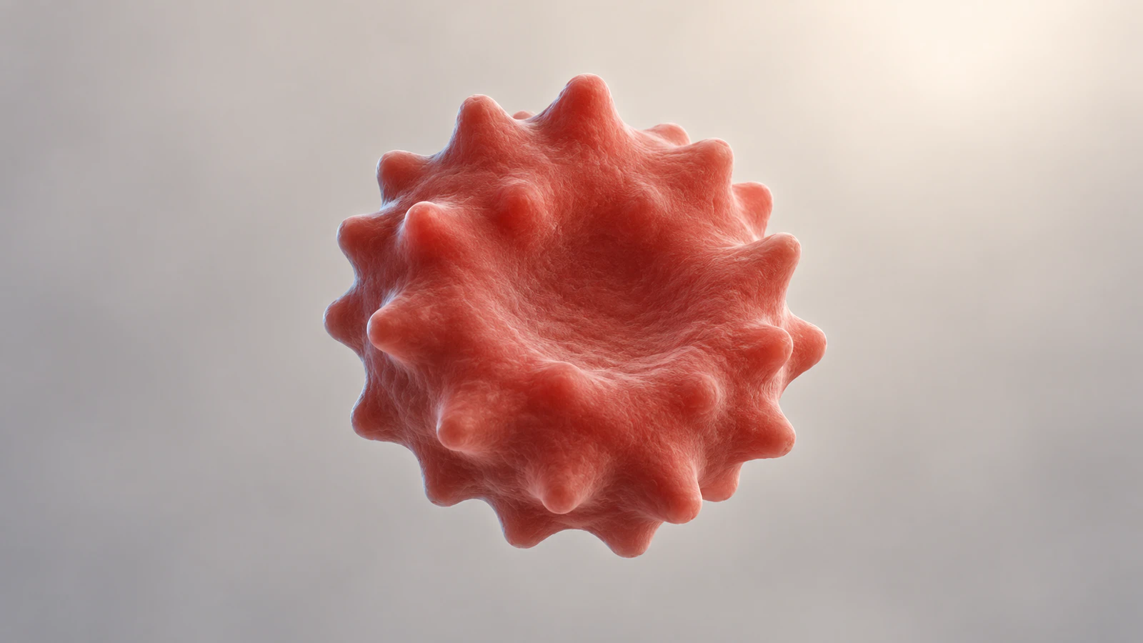
Use the endpoint to show a qualitative consequence, then stop before it looks like experimental proof. The compact, scalloped cell in the final illustration represents a possible shrinkage appearance. Its size cannot be compared numerically with the cover because the images have independent cameras and independently generated geometry.
In your animation, keep the comparison camera fixed and save the initial view. This makes a claimed change easier to inspect. If the project requires a real volume measurement, bring in the relevant data and a defined measurement method rather than estimating volume from the apparent width of a rendered silhouette.
Do not use a cell's appearance alone to diagnose a sample or identify the surrounding solution. An introductory model intentionally leaves out many experimental details. A useful caption names what the image illustrates and what remains unspecified: composition, time, measured volume and exact membrane geometry.
Create the draft in Animiotics
Animiotics lets you start a scientific 3D scene from a written description and refine it with follow-up requests. Begin with one branch of the explanation. The live homepage states that joining is free while AI generation and exports require a paid plan; check the controls available to your account before starting production.
A focused starting direction could be: Create a conceptual red blood cell in a neutral surrounding medium. Keep its membrane intact and the camera fixed. Show a restrained qualitative reduction in volume for a water-loss explanation, then hold the endpoint. Do not add a nucleus, labels, measured values or an automatic burst. This is a proposed prompt to inspect, not a tested result or an accuracy guarantee.
Create the membrane exchange shot separately so you can review it at its own scale. A useful correction request is: Keep the membrane and camera, but show representative water crossings in both directions while retaining the chosen solute on its original side. Review the actual scene after each change. If the model violates the permeability rule, fix that before spending time on lighting.
Test understanding without the labels
Ask a classmate to watch the draft with the condition names hidden. Give them the membrane rule and ask three questions: which way is the net transfer, what happens to the cell and would individual water motion stop at balance? Their explanation should refer to the setup rather than the color palette.
Then change just one assumption in the discussion: suppose the previously retained solute can cross during the interval. Ask whether the original prediction can be reused unchanged. The purpose is to reveal dependence on the membrane, not to solve every possible transport problem in the same video.
Save the scene specification with the reviewed animation. Record the starting condition, permitted crossings, chosen endpoint and any exaggerated timing. When you shorten the animation for a later presentation, this record helps you preserve the setup instead of accidentally keeping only the visually dramatic transformation.
Frequently asked questions
Does water move only one way during osmosis?
No. Distinguish individual crossings from the net transfer. Your animation should make the overall imbalance understandable without implying that every water molecule travels in the same direction.
Does isotonic mean that water stops moving?
No. A stable average cell volume does not require all water exchange to stop. Keep the molecular and whole-cell views conceptually separate.
Can I label a solution from its total solute count alone?
Not for a general cell-volume prediction. Define the cell, membrane permeability and relevant interval. This guide's simple comparison assumes the chosen solute cannot cross during the example.
Should an osmosis animation always show a cell bursting?
No. Choose the endpoint that fits the declared conditions and teaching question. Bursting should not be an automatic visual finale.
Are these images exact aquaporin or red-cell models?
No. They are conceptual illustrations with independent geometry and cameras. Use verified structural or experimental inputs when your explanation requires quantitative or atomic detail.
Try one osmosis scene
Try Animiotics with the water-loss branch first: one intact cell, a fixed camera and a clearly declared membrane rule. Pair the scene with a short exchange shot and ask a classmate to explain the connection. Expand to the other conditions only after that first relationship is clear.