Decide what the glycan must explain
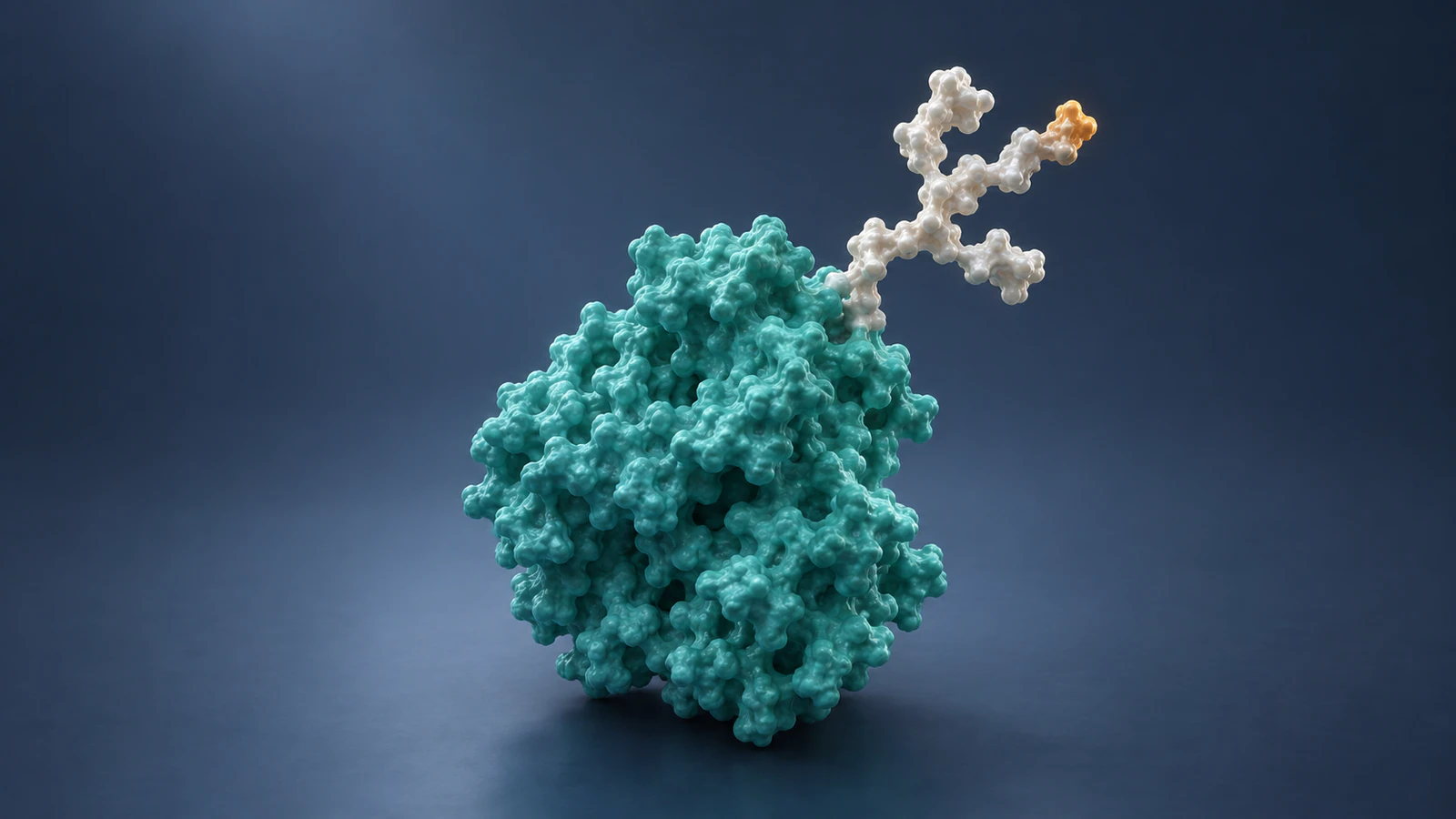
A useful glycan animation separates four questions: where the sugar chain attaches, what it contains, which coordinates are supported and how its movement was chosen. Answer those before adding branches to a protein. Otherwise, a polished scene can make an illustrative decoration look like a fully characterized molecular structure.
For a science creator working in Animiotics, the immediate decision is the level of detail the explanation needs. An overview may only need to distinguish a protein from its attached carbohydrate. A comparison of particular glycoforms needs stronger evidence and a more explicit explanation of what changes between views.
This guide proposes a short evidence record and a six-shot storyboard for making that decision. The worked example is hypothetical, and the images are conceptual illustrations rather than data-derived structures. Use the framework to plan a scene and review its claims before treating it as a finished scientific explanation.
Define the attachment before the shape
A glycan is a chain of sugar building blocks, and a glycoprotein is a protein with carbohydrate attached. Glycans can branch. That branching gives an animator a useful silhouette, but a branch drawn for readability is not enough to identify a particular sugar sequence or linkage. The RCSB carbohydrate guide explains these distinctions.
For common eukaryotic N-linked glycosylation, the attachment is to asparagine, an amino acid often abbreviated Asn. The familiar sequence motif Asn-X-Ser/Thr, where X is not proline, identifies a potential site rather than proving that the site is glycosylated. This is a common motif, not a universal definition of every possible glycan attachment.
The N-Glycans chapter in Essentials of Glycobiology provides that biological context. In a scene review, ask for the source supporting the particular attachment you show. If the assignment is purely conceptual, describe the location as illustrative rather than assigning an invented residue number.
Keep four kinds of evidence separate
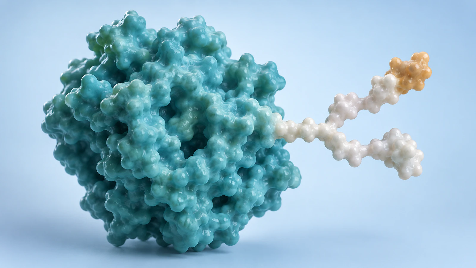
Create one record for each attachment that matters to the story. Do not use a single note saying that the protein is glycosylated as permission to specify every visible branch. Someone reviewing the scene should be able to trace each important visual decision to either a source or an explicitly stated illustrative choice.
The record below is a production tool, not a scoring system for experimental quality. Fill uncertain fields with an actual limit such as composition unspecified. A blank field is too easy to mistake for an overlooked task that the artist should solve by adding detail.
| Field | Record before production | What the image may imply |
|---|---|---|
| Attachment | Protein identity, site and supporting evidence | This chain is connected at this location |
| Composition | Sugar identities and connections, if established | This particular branched glycan is present |
| Coordinates | Source model and any added geometry | These positions have a stated structural basis |
| Motion | Trajectory source or illustrative timing | Movement comes from data or editorial animation |
Read the structure without filling every gap
A deposited structure may contain only part of a glycan. Missing carbohydrate coordinates do not by themselves establish that the biological molecule lacks that carbohydrate. The RCSB carbohydrate guide specifically notes that an entry may include only the first resolved sugar, with other portions unavailable in the coordinate model.
Inspect the entry before deciding that a short chain needs extending. The RCSB Structure Summary documentation describes oligosaccharide names, chain identifiers and interaction views. A single modeled sugar can be listed among ligands rather than in the oligosaccharide section, so check both places.
Save the entry version and the coordinate file you actually use. In your production notes, separate geometry retained from that file from anything supplied by another model or drawn conceptually. For the related protein problem, see how to show missing residues honestly. Neither a tidy silhouette nor an empty patch should silently resolve an uncertainty.
Choose a representation for the question
Choose a molecular surface when the reader needs a spatial overview, a validated chemical representation when the connections are the point, or a clearly described schematic when the story is about organization. Do not switch between these treatments without explaining the change. A viewer may otherwise read a change in drawing style as a change in the molecule.
The Symbol Nomenclature for Glycans, or SNFG, is a defined convention. If you use it, follow its symbol rules and provide the information needed to interpret the drawing. A custom pearl-and-amber palette is an art direction choice, not an alternative SNFG encoding.
Our illustrations use continuous surface-like envelopes to make attachment and branching readable. They do not specify sugar identities, stereochemistry or exact bonds. When your teaching objective requires those details, replace the conceptual geometry with a reviewed representation rather than asking the caption to excuse an inaccurate chemical diagram.
Show variation without inventing a population
Glycoforms are versions of a glycoprotein that differ in their glycan complement. The N-Glycans chapter describes how glycan variation can occur at a site and depend on the biological production context. One appealing branched model should therefore not automatically stand for every molecule in a sample.
For a qualitative comparison, show selected examples and say that they are examples. Repeating one version many times can suggest its abundance even when no proportions are stated. If you have sample-specific measurements, cite them and decide how the display represents them; otherwise, avoid countable crowds that look like population data.
Keep the protein view and scale matched when the glycan is the comparison variable. Separate the examples enough that they cannot be mistaken for an interacting pair. Do not animate one into the other unless transformation is the actual claim and the sequence has its own biological support.
Work through a two-example scene
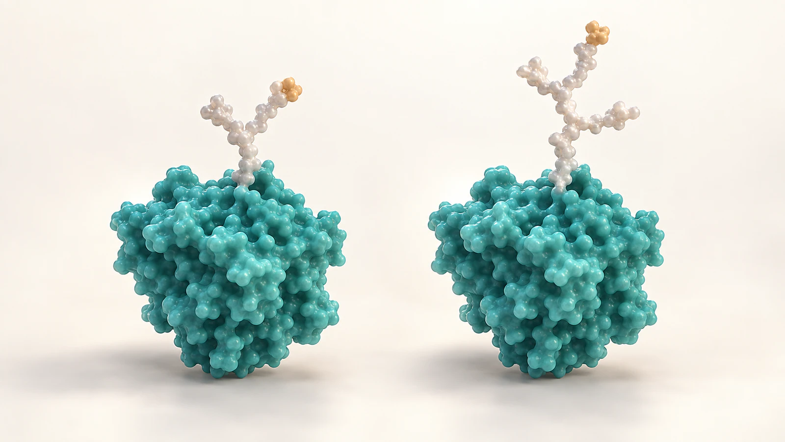
Suppose your task is to explain that an attached carbohydrate can vary while keeping the protein recognizable. You have permission to use a generic glycoprotein illustration but no sample-specific site map, glycan sequence or trajectory. The responsible deliverable is a conceptual comparison, not a reconstruction of an unnamed experiment.
Write the four-field record first: illustrative attachment location, unspecified sugar composition, artist-created geometry and no measured motion. Choose two visibly different branch arrangements while retaining the same protein shape. Call them selected conceptual examples in the narration or caption, and avoid language such as dominant form or fully occupied.
Use a cut between examples or place them beside one another. A growing branch would tell a different story about construction, while a dissolving branch could imply removal. The scene succeeds when a reviewer can describe the intended variation without also reporting a reaction, a frequency or an exact chemical structure that you never meant to show.
Plan six shots with clear boundaries
Treat this sequence as a storyboard proposal for the hypothetical comparison, not a description of a measured process. Each shot has one job and one limit. Set screen duration for readability, and keep that editorial duration separate from biological time.
Before rendering the sequence, read the shot descriptions aloud without showing any images. If the words promise only a comparison but the proposed movement depicts a reaction, change the movement. This is cheaper and clearer than trying to repair the message with a disclaimer after the animation is finished.
| Shot | Purpose | Review boundary |
|---|---|---|
| 1. Establish | Show the complete protein and attached chain | Do not imply a named structure |
| 2. Locate | Move closer to the attachment | Do not invent a residue identity |
| 3. Follow | Let the eye trace the branch arrangement | Do not imply sugar-by-sugar synthesis |
| 4. Compare | Introduce a second selected example | Do not suggest population proportions |
| 5. Hold | Keep both examples at matched scale | Do not turn framing into a size claim |
| 6. Resolve | Return to one readable overview | State what is illustrative and what remains unspecified |
Make the attachment readable at small size
Check the frame as a small thumbnail before adding more detail. The protein body, the attached chain and the connection should remain distinguishable. If the attachment disappears in shadow, adjust the camera or light before thickening the chain; changing geometry to fix composition can create a new scientific claim.
Use a quiet background and a restrained accent to guide attention. In a sequence, keep the protein and carbohydrate treatments consistent so the viewer does not need to relearn their identities at each cut. The molecular color continuity guide offers a practical way to record those choices.
Inspect the root and every branch endpoint at full size as well. Look for a chain that appears detached, passes through the protein or merges with another branch accidentally. A pleasing overview does not compensate for a connection that stops making sense when the reader looks closely.
Review the claim carried by the frame
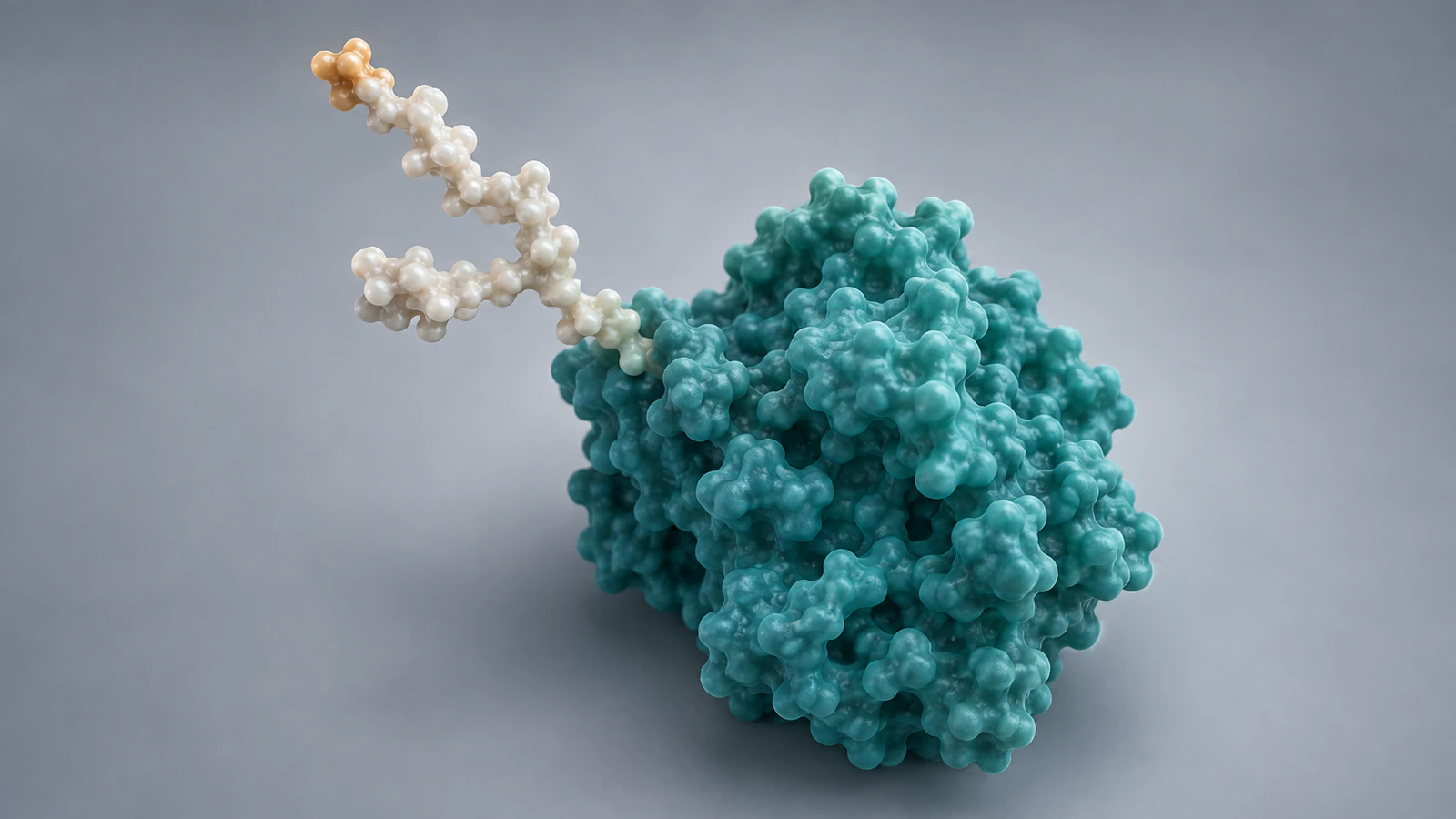
Ask a colleague to describe the image before reading your caption. Listen for unintended conclusions: a specific glycan identity, a measured size difference, a reaction step or a claim that one version is more common. Revise the scene when its strongest visual message goes beyond the evidence record.
Write the caption to identify the representation and explain its useful limit. For example: conceptual comparison of two branch arrangements; the geometry does not specify sugar identities or sample frequencies. This tells the reader what they can learn without pretending the illustration provides data it does not contain.
Keep a final handoff note with the source links, model versions, four-field records and approved captions. If someone later requests a binding or shielding sequence, reopen the evidence review rather than treating the existing glycan as proof of that effect. A new mechanistic claim needs its own support.
FAQ
Does a potential N-glycosylation site prove a glycan is present?
No. A sequence motif identifies a potential site; show a specific attachment as established only when the relevant evidence supports it.
Does a short glycan in a structure mean the full molecule has only that chain?
Not necessarily. The coordinate model may contain only the portion that could be modeled from the experimental data, so inspect the entry and supporting study.
Can two glycan examples represent a sample distribution?
Two selected examples alone do not establish frequencies. Describe them as qualitative examples unless a measured distribution supports the way you display proportions.
Are the images here chemical structures or SNFG diagrams?
No. They are independent conceptual CGI illustrations with surface-like envelopes. The colors and branches do not encode exact sugar identities, chemical linkages or data-derived geometry.
Can I add gentle movement for clarity?
You can use illustrative movement if its status is clear and it preserves the intended attachment. Do not present artistic motion or screen duration as a measured trajectory or biological timescale.
Try one glycoprotein explanation
Try Animiotics with a focused scene: one recognizable protein, one attached carbohydrate and a caption that says which details are illustrative. Use the four-field record to decide what the animation should communicate before expanding it into a comparison. Review the attachment in both a close view and the final small frame.
Keep the first goal specific: help a viewer distinguish the attached chain from the protein without inferring an exact glycoform. Visit the homepage to explore Animiotics and its current access options.