Follow the phosphate, then explain the effect
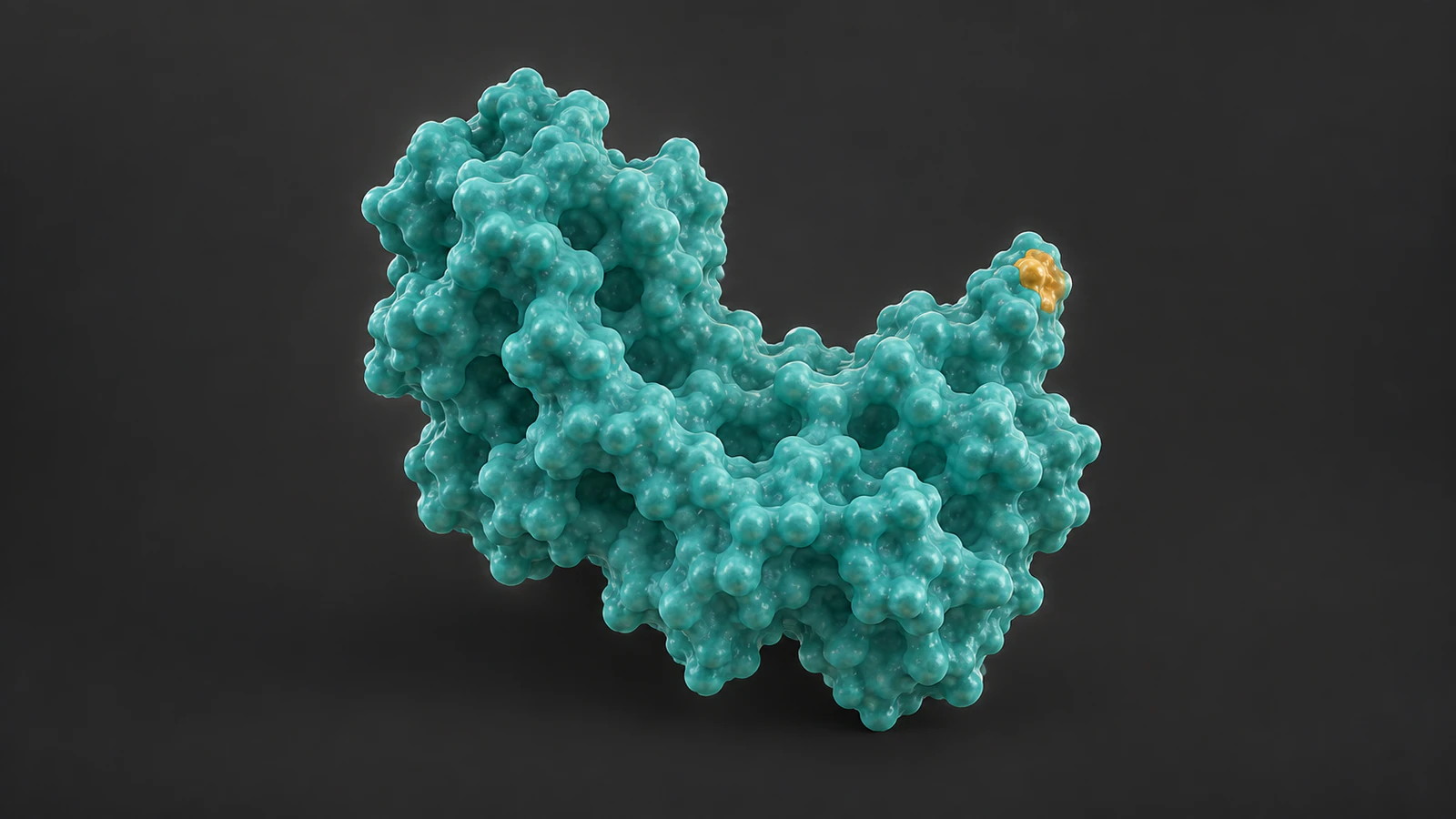
A protein phosphorylation animation should show a phosphate added at a defined site, identify its donor and keep the resulting modification separate from any claim about protein activity. If the story includes removal, introduce a phosphatase and a distinct reaction. Making the entire protein light up skips the most useful explanation.
For a scientist or creator working in Animiotics, the first deliverable can be a short sequence that answers three questions: what changes chemically, which actors cause that change and what consequence the evidence supports. These decisions belong in the storyboard before camera movement or surface detail makes the sequence expensive to revise.
This guide uses the common serine, threonine and tyrosine phosphorylation scheme as its scope. The worked example is deliberately unnamed: it teaches reaction accounting without presenting an invented protein shape, residue number or activity measurement as a real experimental result.
Define the chemical scope
A residue is an amino acid unit within a protein chain. In the conventional scheme described here, a protein kinase transfers the terminal phosphate group from adenosine triphosphate, or ATP, to a hydroxyl group on an appropriate side chain. Adenosine diphosphate, or ADP, is the nucleotide product. This is a covalent modification, not merely a small molecule sitting in a pocket. NCBI: Protein Function.
Write the chosen residue type in the working notes even if the public scene stays at surface level. For a named target, also record the organism, protein form and numbering convention. A technically impressive close-up becomes misleading if the highlighted site belongs to another isoform or the viewer cannot tell which chain carries it.
Limit this first sequence to one selected site. Do not imply that all phosphorylation uses these three amino acids or that every exposed hydroxyl is a suitable substrate. A narrow scope lets the animation explain one event clearly while leaving other chemistries and multisite regulation for a separate, properly sourced story.
Make a reaction-accounting worksheet
Use one row per reaction rather than one row per camera shot. A shot can hide a product behind the enzyme, but that product should still have an explicit place in the production notes. This distinction catches disappearing ATP and mysteriously regenerating ATP before either error becomes part of the animation.
The worksheet below is a planning convention, not a balanced chemical equation. It omits protonation details, metal coordination and atom-level intermediates. Add those only when your specific scientific question requires them and suitable evidence is available. The practical check is whether every emphasized change has an identified starting state and destination.
| Record | Kinase scene | Phosphatase scene |
|---|---|---|
| Protein state | Selected hydroxyl site before modification | Same selected site carrying phosphate |
| Reaction partner | ATP supplies the transferred group | Water participates in hydrolysis |
| Displayed result | Phosphorylated site and ADP | Dephosphorylated site and inorganic phosphate |
| Catalyst identity | Named or explicitly conceptual kinase | Separate named or conceptual phosphatase |
| Effect on activity | State only what evidence establishes | Do not assume a universal on/off reversal |
Keep the catalytic encounter readable
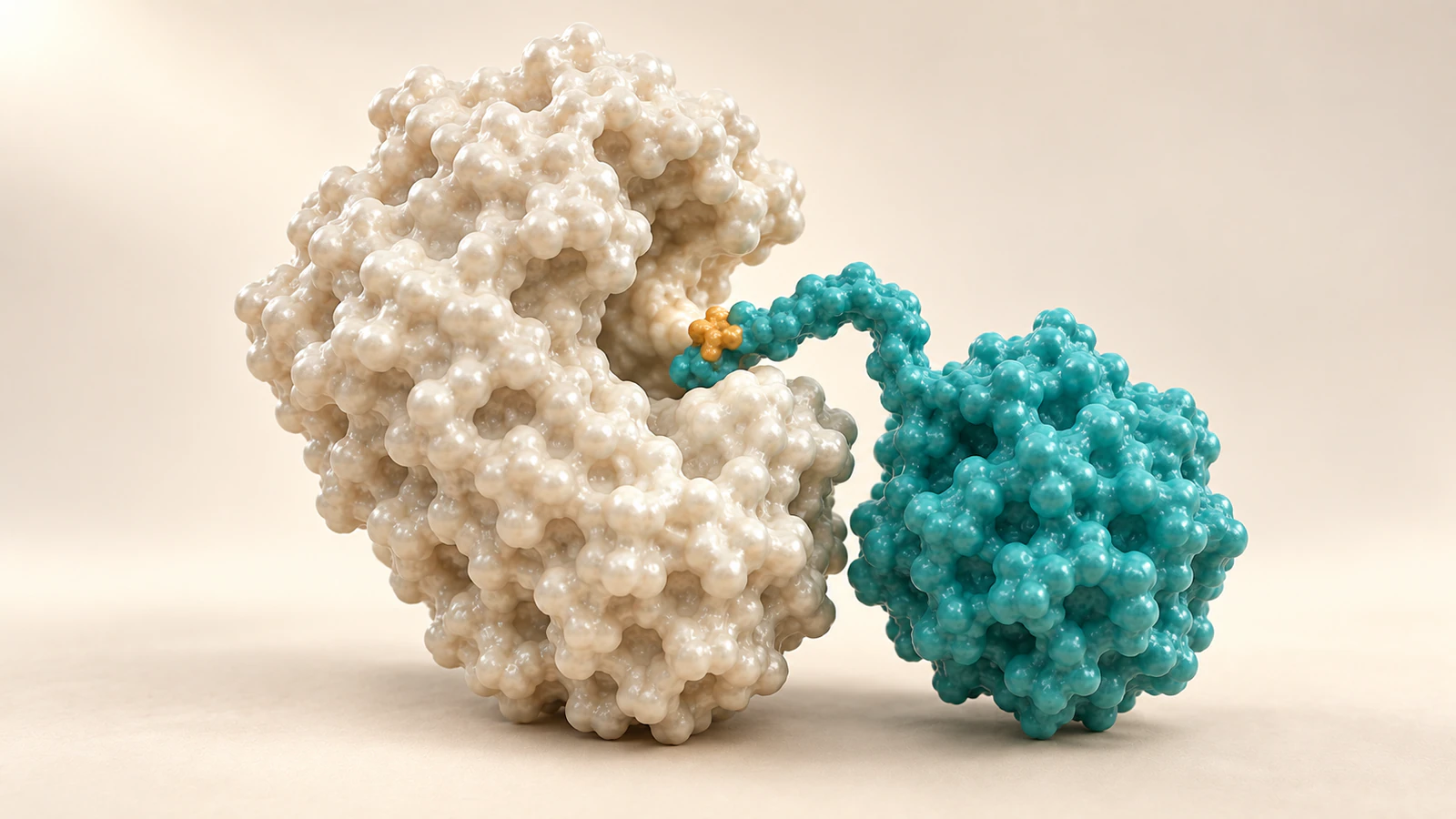
The substrate is the molecule acted on by an enzyme. At the protein scale, stage the encounter so the viewer can distinguish the kinase from the substrate and follow the selected region into the catalytic cleft. Avoid merging the two surfaces into a single mass or making the substrate pass through an opaque enzyme.
A close-up can explain access and orientation while remaining openly schematic. The RCSB discussion of protein kinase A illustrates why ATP positioning and metal ions matter when examining actual catalytic structures. Those details require structural evidence; an attractive surface model cannot supply them. RCSB PDB-101: PKA.
Choose whether the scene promises chemistry or spatial context. For chemistry, use validated atom identities, connectivity and the relevant reaction model. For context, show the encounter and clearly describe omitted small molecules in the caption or narration. Do not add an arbitrary atom cluster simply to make a surface-level shot look more technical.
Avoid turning modification into activation
Phosphorylation can activate or inhibit protein function. The direction depends on the particular protein, site and biological setting. A kinase adding a group therefore does not justify an automatic green glow, an opening active site or a downstream burst of signaling. NCBI: Regulation of Protein Function.
Separate the visual statement “this site is modified” from the statement “this protein is more active.” The first can be represented with a localized marker. The second needs an identified activity readout and the conditions under which it was observed. If your evidence only establishes modification, end the mechanistic beat there.
A phosphate can influence conformation or create a recognition feature for another protein, as the PDB-101 explanation describes. Treat these as possible consequences to investigate, not stock motions to apply to every model. A restrained hold on the modified site can communicate the evidence more accurately than an unsupported dramatic transformation.
Separate the cast before animating
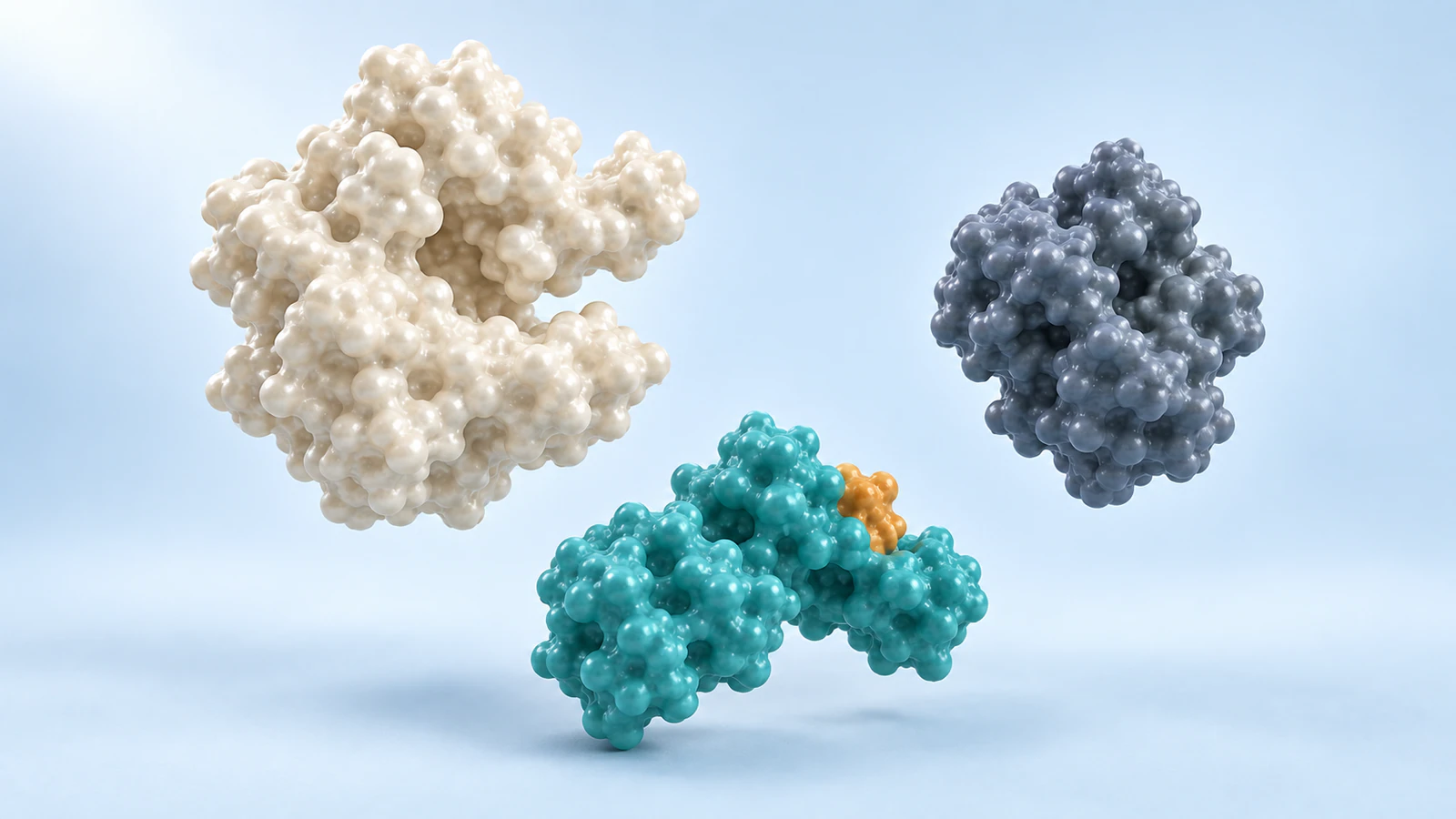
Prepare a simple cast view with the substrate, kinase and phosphatase visibly distinct. Give each actor a stable name in the working file and a silhouette that survives a change in camera angle. Keep the substrate identity consistent through both reactions; the enzymes should not quietly exchange roles during a cut.
Use color to reinforce that identity. A small amber marker can indicate the selected modification while the rest of the substrate retains its original material. Explain any enlargement of the marker: visibility at presentation size is an editorial choice, not evidence that phosphate occupies a large fraction of the protein.
The accompanying cast illustration uses independent conceptual surfaces to make the roles legible. Its sizes and spacing are not molecular measurements. For a continuous production sequence, reuse the same reviewed substrate geometry across shots. The method in molecular color continuity helps keep identity separate from state.
Build a six-shot worked example
Imagine an unnamed substrate with one selected serine site. The brief is to explain addition and removal, with no asserted change in activity. Keep the camera on the same side of the substrate during both encounters so the audience can recognize the site without relying on a repeated label.
Treat these six shots as an editorial sequence, not a measured trajectory. The holds can be lengthened for teaching, and transitions can be simplified, provided the viewer is told that molecular timing is illustrative. Use the worksheet to decide which products remain visible and which are explicitly accounted for off-screen.
- Shot 1: Establish the unmodified substrate and selected region. Identify the kinase separately, leaving enough space to recognize both silhouettes.
- Shot 2: Frame the catalytic encounter. Explain ATP as the donor through a validated chemical view or clear narration; retain the substrate boundary.
- Shot 3: Show the selected site as phosphorylated and account for ADP. Hold the composition long enough to inspect the localized change.
- Shot 4: Separate the substrate from the kinase. Preserve its identity and modification marker without adding an unsupported activity effect.
- Shot 5: Introduce the distinct phosphatase at the selected site. Explain hydrolysis rather than reversing the earlier transfer shot.
- Shot 6: Show the dephosphorylated substrate and account for released inorganic phosphate. Conclude with the modification state that the sequence actually demonstrated.
Give removal its own reaction
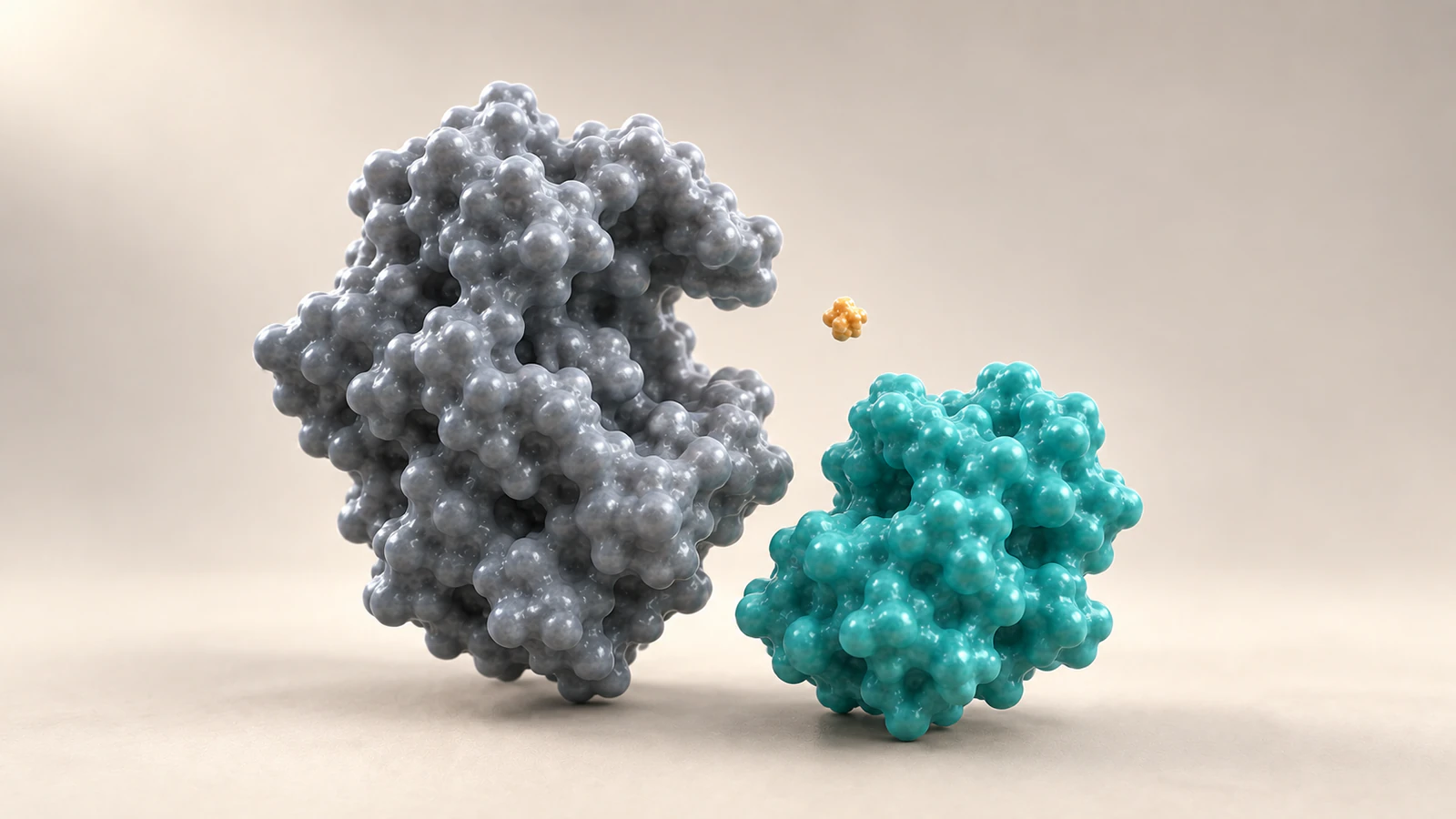
Protein phosphatases catalyze hydrolysis of phosphorylated residues. In this scheme, removal yields a dephosphorylated site and inorganic phosphate. The animation should not show the phosphate returning to ADP to rebuild ATP as though the phosphatase were simply running the kinase process backward. NCBI: Regulation of Protein Function.
Changing only the direction of a motion path is especially tempting when building a looping scene. Resist that shortcut. Make a separate removal beat with its own catalyst, starting state and product accounting. If the public loop returns to the opening frame, use an editorial transition rather than depicting unsupported ATP regeneration.
Do not make the phosphatase erase every modification on the protein unless that scope is supported. Keep the selected site visible through removal, then inspect the final frame against the initial substrate record. The visual comparison should concern that site, not imply that every molecular property or cellular effect has returned to its starting value.
Match the image to the evidence
A conceptual molecular surface can communicate the location of an encounter. It cannot establish a residue identity, hydrogen-bond network or transition state. If those details are central, move to a reviewed structural representation and identify the data source. See protein surface versus ribbon for choosing a view that answers the actual question.
A structure and an activity assay also answer different questions. Keep the evidence beside the claim it supports rather than letting a detailed molecular image stand in for every type of result. A caption can state that a site is illustrated schematically while a separate cited result describes the observed functional consequence.
For a named project, ask the scientific reviewer to approve the residue assignment and interpretation before approving polish. Record unresolved issues explicitly: uncertain site placement, missing local coordinates or unavailable product-state structure. Choose a conceptual treatment when that is the honest level of detail, and explain the boundary without burying it in fine print.
Review the sequence without narration
Run the first review with the sound off. Ask the reviewer to point to the substrate, identify the reaction site and explain what changed between the opening and final frames. If the answer is simply “the protein became active,” your visual language may be making a stronger claim than the sequence supports.
Then restore narration and compare it with the worksheet. Check that ATP and ADP are distinguished, the removal product is accounted for and the two catalysts remain recognizable. Inspect still frames at slide size as well as full resolution: a marker that vanishes at small size or looks detached can change the meaning of the shot.
- Chemical scope: one selected site, explicit donor and products, no accidental ATP regeneration.
- Identity: substrate stays recognizable; kinase and phosphatase remain separate actors.
- Evidence: every motion or activity consequence is supported or identified as illustrative.
- Presentation: captions explain omissions, marker enlargement and conceptual geometry.
FAQ
Does phosphorylation always activate a protein?
No. The effect depends on the protein, site and context. Show the modification separately from activation or inhibition, and only add a functional consequence when the evidence supports it.
Should ATP appear in every shot?
No. A surface-level sequence can omit small molecules visually if narration or captions explain the donor and products. An atom-level transfer scene requires a chemically reviewed representation rather than an arbitrary decorative ATP shape.
Can dephosphorylation reuse the kinase animation backward?
That would misrepresent the reaction described here. Use a distinct phosphatase scene and account for hydrolysis and inorganic phosphate instead of showing ATP being rebuilt.
Can a phosphate marker be enlarged for readability?
Yes, as an explicitly illustrative device. Keep it localized to the selected site and explain that its displayed size and color are not molecular measurements.
Do these images depict a real protein structure?
No. They are conceptual CGI illustrations of scene roles and modification states. Their geometry, spacing and amber markers are not derived from a named structure or experimental trajectory.
Try a phosphorylation sequence in Animiotics
Try Animiotics with a short animation of one substrate passing through a kinase encounter and a separate phosphatase encounter. Use the six-shot example as your starting plan, then review the selected site, donor, products and supported consequence before extending the sequence into a larger pathway story.
Keep the reaction worksheet alongside your references while you create. A useful first result is a sequence whose chemical meaning survives each camera cut and whose captions clearly distinguish the conceptual scene from the evidence.