Align first, then inspect the fit
To align two protein structures in ChimeraX, open both coordinate files and run Matchmaker with one chain as the moving structure and the other as the reference. For the example below, the command is matchmaker #2/A to #1/A pairing ss showAlignment true. It superimposes chain A of 4AKE onto chain A of 1AKE and displays their sequence alignment.
Read the Log before judging the overlay. The fitted subset can agree closely while other regions remain far apart. Record the chain choices, fitting settings and atom-pair counts with the image; an attractive superposition alone does not establish a biological motion pathway.
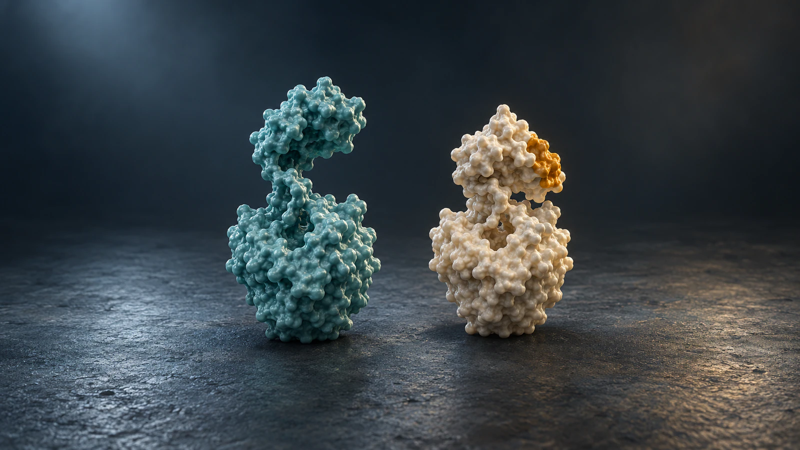
This guide adds a concrete alignment and reporting procedure to our ChimeraX movie tutorial. Its four illustrations are conceptual CGI, not coordinate-derived depictions of the example structures.
Choose a defensible comparison
The worked pair is Escherichia coli adenylate kinase, an enzyme involved in adenine nucleotide interconversion. 1AKE contains the inhibitor Ap5A, which mimics the two substrates. 4AKE is an unligated structure described in a study of conformational differences in the same enzyme.
Both entries list protein chains A and B with 214 residues per chain and no mutations. We deliberately compare chain A from each entry. This is a single-chain structural comparison, not a comparison of their complete biological assemblies.
That scope matters: the current RCSB records give different assembly descriptions for these entries. Selecting matching chain letters is a reproducible choice here, but letters do not establish equivalence in arbitrary structures. Check sequence, organism, construct and ligand context before reusing this procedure with another pair.
Keep the entry pages and source files with the project. If your question concerns a functional complex rather than one polypeptide chain, first use the biological assembly guide to decide which molecular unit belongs in the comparison.
Set up a clean session
Use UCSF ChimeraX, with the Command Line and Log available, and internet access for fetching the PDB entries. Save any existing work and start a separate empty session so the first two structures receive model numbers #1 and #2. Model numbers identify loaded objects; /A specifies chain A.
The loading, representation-setting, fitting and session-save commands below were executed in ChimeraX 1.9 on Windows in headless mode on October 8, 2026. Current official documentation and the ChimeraX 1.12 production-release notice were also checked. Interactive framing, PNG rendering and the alignment window were not tested in that headless run.
Enter these commands one at a time. If your session already contains models, inspect the Model Panel and replace the numbers throughout rather than assuming they match the example.
- open 1ake
- open 4ake
Show only the chains being compared
Remove atom clutter and the second protein chain from the visible cartoon representation before fitting. The following sequence leaves the chosen chains visible and gives each structure one persistent color. It changes display settings, not which coordinates exist in the loaded files.
Keeping both models is useful: you can revisit ligands or the other chain later without fetching the entries again. Here, teal identifies 1AKE and gold identifies 4AKE. Those colors are presentation choices and carry no confidence, energy or activity meaning.
- hide atoms
- hide cartoons
- cartoon #1/A
- cartoon #2/A
- color #1/A teal target c
- color #2/A gold target c
Run the chain-specific alignment
Run matchmaker #2/A to #1/A pairing ss showAlignment true, followed by view #1,2/A. The first command fits the selected chain from model #2 onto the reference chain in model #1. The second frames the specified chains together without defining a new structural fit.
The Matchmaker documentation distinguishes sequence correspondence from spatial fitting. For proteins, the fitting points are alpha carbons, the backbone atoms named CA. The pairing ss option explicitly supplies the two chains rather than asking the tool to choose a best-scoring chain pair.
The expected result is an overlay with better agreement in some regions and visible separation elsewhere. In the graphical application, the sequence alignment helps inspect residue correspondence. Do not drag one model until it looks better after fitting; that would change the spatial comparison you are trying to report.
Our local run completed successfully and created a saved session. It reported 112 retained atom pairs with an RMSD of 1.083 Å, and 8.240 Å across all 214 aligned pairs at that fitted placement. These are results of this specific command and version, not universal reference values for every comparison of the entries.
Report the pairs behind the RMSD
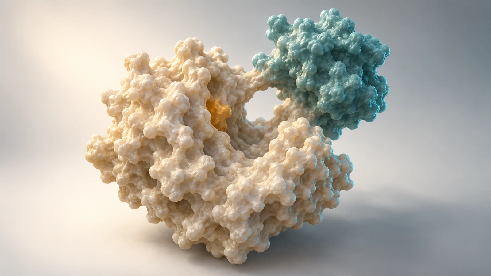
Root-mean-square deviation, or RMSD, summarizes distances between corresponding points. The ångström, written Å, is a length unit equal to one ten-billionth of a metre. A reported deviation needs the atom selection and fitting method to be interpretable.
Matchmaker normally iterates the fit and removes more distant pairs using its cutoff rule. This explains how our retained subset can have a low deviation while the full correspondence has a much larger one. The larger value describes all aligned pairs after the subset-based fit; it is not the result of a separate best fit to all pairs.
For a deliberate all-pairs comparison, run matchmaker #2/A to #1/A pairing ss cutoffDistance none. This turns off pruning and changes the fit. Save the original result first, then record the new output separately. Neither setting should be chosen solely because it produces the smaller headline number.
The practical question is which comparison answers your figure caption. If you need the position of a mobile region relative to a specific domain, justify that reference region from structural evidence. Do not present an automatically retained subset as a biologically validated domain boundary.
Keep a comparison record
A useful handoff includes enough information for a colleague to recreate the placement and identify what is absent from the picture. Use the following record alongside your figure or movie project. Copy numerical results from your own Log rather than from this article.
For this example, a concise caption could state that chain A of 4AKE was superimposed on chain A of 1AKE using ChimeraX Matchmaker with iterative fitting, with teal and gold identifying the two entries. Add the retained-pair count and both deviations if the caption discusses numerical agreement.
| Record | What to retain |
|---|---|
| Inputs | PDB identifiers, coordinate downloads, chain choices and ligand context |
| Execution | ChimeraX version, exact command and reference-versus-moving model |
| Correspondence | Sequence alignment, gaps and any residue restrictions |
| Fit | Pruning setting, retained-pair count and reported RMSD values |
| Display | Hidden chains, hidden ligands, colors and chosen camera |
| Interpretation | What the overlay supports and what remains unmeasured |
Save a figure you can reconstruct
Save the session with save ~/Desktop/ake-comparison.cxs. Choose an existing writable folder if Desktop is elsewhere on your system. A ChimeraX session preserves the working setup for later inspection; keep the source entries and command record alongside it.
For a still image, the documented command is save ~/Desktop/ake-comparison.png width 2400 height 1600 supersample 3. These pixel dimensions are an illustrative output choice, not a journal requirement. Inspect the preview framing before export and open the saved image afterward to check that the entire comparison is visible.
The PNG command is documentation-checked, not a claimed local render test. Use the transparent-background export guide if the figure needs to sit on another slide background. Preserve the same colors and legend when moving between a still and a movie.
Troubleshoot the actual mismatch
When a fit fails or looks confusing, inspect the inputs before changing visual styling. A clearer silhouette cannot correct a wrong chain pairing. Work through one issue at a time and keep the last interpretable session.
| Symptom | Check | Next action |
|---|---|---|
| No atoms or chains match | Model numbers, chain IDs and polymer type | Inspect the loaded models and correct the specifications |
| Two copies clutter the view | Extra cartoons or visible atomic representations | Repeat the display commands for the chosen chains |
| Low RMSD, large visible separation | Retained pairs versus all aligned pairs | Read both values and inspect the pruned correspondence |
| Overlay changes after manual dragging | Relative model movement after fitting | Rerun the same command before saving |
| Need a specified atom subset | Whether sequence-based matching answers the question | Use the documented align command only with justified atom correspondence |
| Saved image crops a domain | Camera framing and output aspect ratio | Reframe both selected chains and inspect the saved file |
Separate comparison from an animated pathway
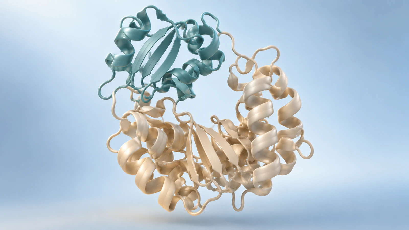
A superposition places structural models in a shared frame. It does not measure the order, speed or probability of movements between them. Even a convincing open-and-closed pair leaves those questions unanswered unless other evidence supplies them.
For a teaching sequence, first show the reference, then reveal the comparison in the same camera view, then pause on the separated region. A cut between endpoints is often sufficient. If you introduce an interpolated transition, identify it as an illustrative transition and retain the two experimentally derived endpoints as distinct evidence.
ChimeraX provides a separate morph command that constructs intermediate coordinate sets. Our morph-versus-simulation guide explains why those intermediate frames should not be described as a measured trajectory. Keep any invented playback duration out of claims about molecular timescales.
A reproducible conceptual scene plan for Animiotics is: hold a simplified enzyme with its cleft open, keep the core and camera fixed, move only the designated lobe toward the core, then hold the alternative state. Review the connection between lobe and core frame by frame. This proposed brief has not been generated or validated as a molecular reconstruction.
FAQ
Does Matchmaker compare every atom?
No. Protein fitting uses one alpha-carbon point per aligned residue, and iterative fitting can prune pairs. Inspect the correspondence and reported pair counts before describing the result as a whole-protein comparison.
Which structure moves in the command?
In matchmaker #2/A to #1/A, model #2 is fitted to the reference in model #1. Keep that direction in the methods note, especially if other objects are positioned relative to one model.
Can I compare different proteins?
Matchmaker supports sequence-based comparison, but a completed fit does not establish biological equivalence. Check sequence alignment, domain composition and the scientific reason for comparing the selected structures.
Is a low RMSD proof of the same conformation?
A low value can describe only a retained subset. Our tested example had substantially different deviations for retained and all aligned pairs, so the selection is part of the result.
Can Animiotics replace this alignment calculation?
This guide uses ChimeraX for structural superposition and numerical reporting. Animiotics can support a separate explanatory animation, which still needs review against the coordinate evidence and the intended biological claim.
Try an enzyme-state explanation
Use the comparison to write a precise animation brief for Animiotics: identify the stable core, the region that changes position and the evidence boundary. Start with the short open-cleft scene above, then inspect whether the intended movement remains clear from a fixed camera.
The current product supports creating an editable 3D scene from a written description and refining its presentation. Signing up and the manual editor are free; AI generation and exports require a paid plan. A generated teaching scene should be reviewed as an illustration, not accepted as the fitted coordinates or a validated transition path.