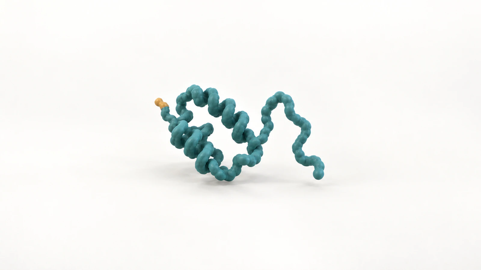
A changed fold or a cut chain
Protein denaturation changes a protein’s folded structure without inherently cutting its polypeptide backbone. Protein degradation breaks that backbone into smaller products through peptide-bond cleavage. In an animation, the decisive difference is therefore continuity: an unfolded chain can remain one connected object, while cleavage creates new chain ends.
That distinction is easy to lose when a compact protein becomes a cloud of particles. The viewer may read the same effect as unfolding, degradation or simple disappearance. A clear sequence shows what changes in shape and what changes in connectivity as separate events.
This guide develops a conceptual example you can plan in Animiotics: one protein unfolds in one branch of the story and is cleaved in another. A six-shot storyboard and a short continuity record make the distinction reviewable. The example is an editorial plan, not a measured trajectory or a claim that an AI-generated animation has been scientifically validated.
What denaturation preserves
A polypeptide is a chain of amino acids joined by peptide bonds. Its primary structure is the amino-acid sequence; higher levels of structure describe how that chain folds and, in some proteins, how multiple chains assemble. A folded shape and an intact sequence are different properties.
The OpenStax protein chapter describes denaturation caused by changes such as temperature, pH or chemical conditions while the primary sequence is retained. For a basic unfolding illustration, keep the backbone connected as its compact organization changes. Do not add a break merely to make the motion more dramatic.
Use an irregular, partly extended shape rather than a perfectly straight rope. The drawing should communicate loss of the original fold without pretending that every unfolded molecule has one prescribed geometry. Avoid a temperature label unless the named protein, conditions and evidence justify that value.
What degradation changes
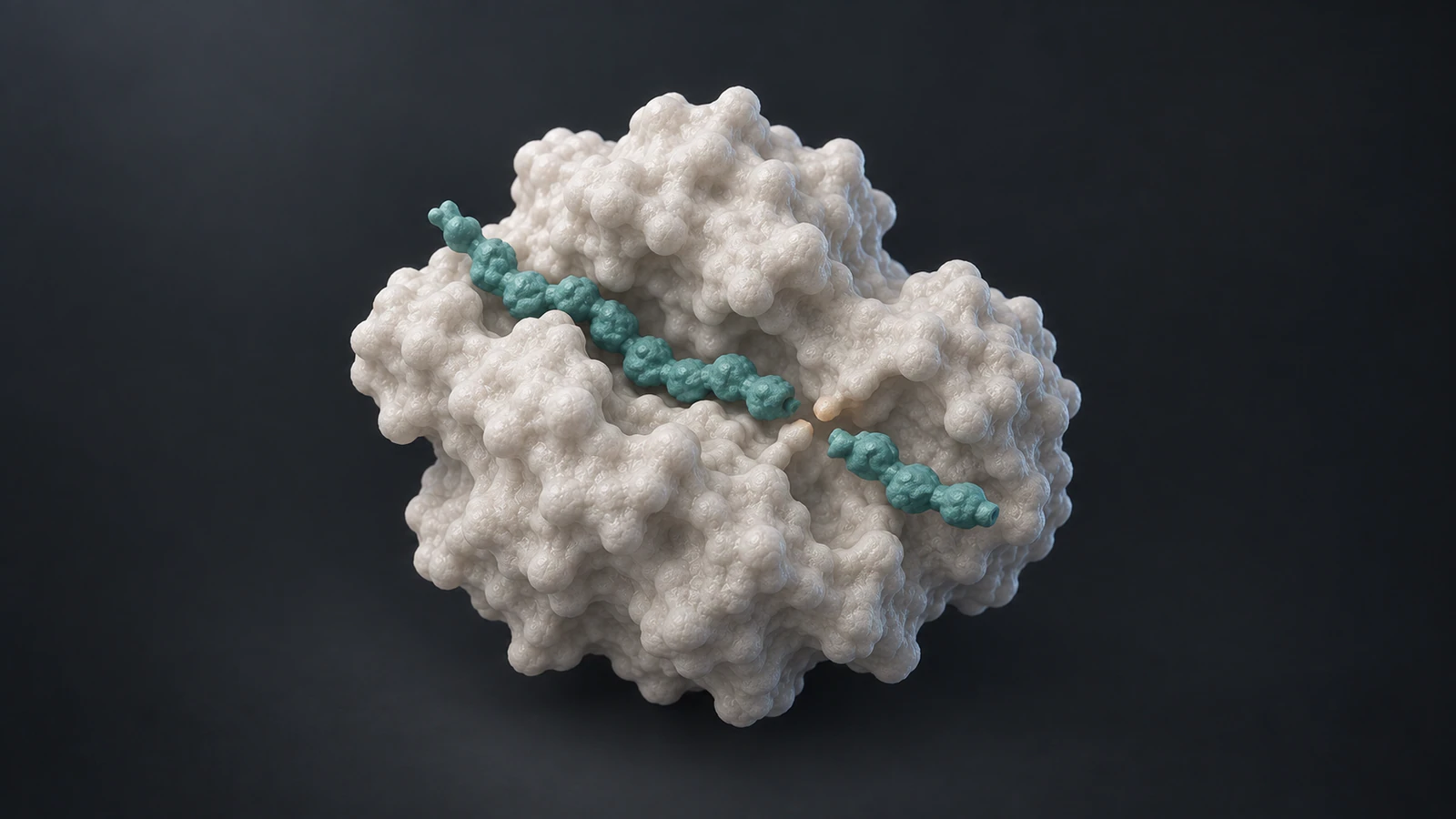
Proteolysis means peptide-bond cleavage. It can produce shorter peptide chains; degradation can involve further processing of those products. A single cut illustrates cleavage, but it should not be presented as the complete breakdown of every protein into free amino acids.
RCSB PDB-101’s proteasome overview describes a regulated molecular machine that can unfold substrates and feed them into an internal protein-cutting core. This is a useful example because unfolding and cutting appear within one pathway while remaining different operations. The proteasome is not the only route by which proteins are degraded.
If the lesson concerns the general distinction, a simplified protease and one visible cleavage are enough. If it concerns proteasomal degradation, add the relevant recognition and processing stages from sources for that system. Do not turn a generic enzyme shape into a supposedly exact proteasome simply by giving it a barrel silhouette.
Keep aggregation separate
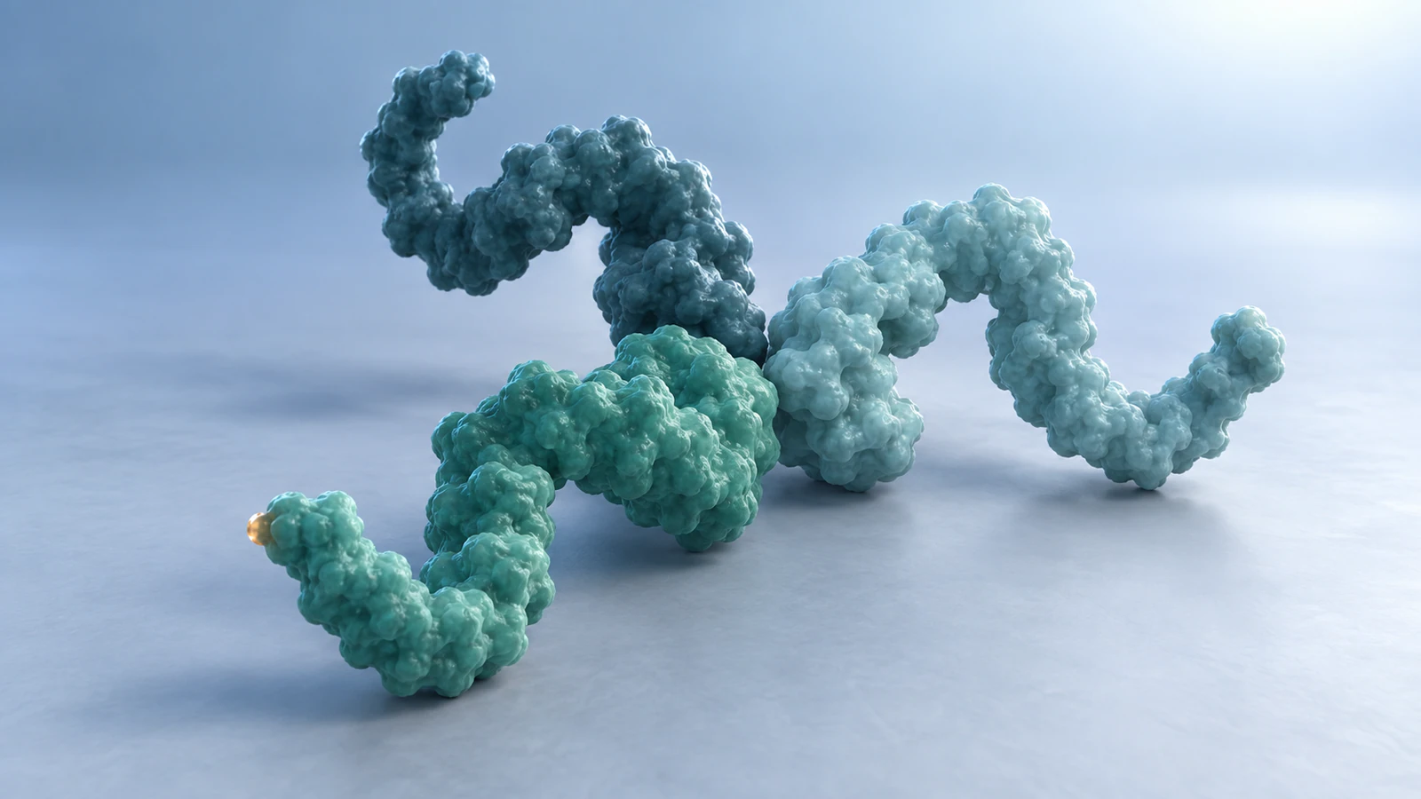
Aggregation is association between protein molecules, not a synonym for cutting a chain. Misfolded proteins can expose regions that favor association with other proteins, as discussed in RCSB’s chaperone overview. Molecular chaperones can assist folding and protect vulnerable regions, but their presence does not establish that every damaged protein will recover.
For an aggregation scene, introduce several molecules before they approach each other. Preserve their separate identities and make the contact regions readable. A group of chains should not silently fuse into one new backbone or become fragments because the camera pulls away.
Treat aggregation as a possible additional branch when it serves the lesson. Do not make it an obligatory intermediate between denaturation and degradation. In a short teaching animation, a caption explaining that this branch is omitted may be more helpful than adding an unsupported chain of biological events.
Write a continuity record
Before animating, record four things for each shot: the chain identity, its folded state, its backbone connections and the evidence being represented. This gives the scientific reviewer something more precise to inspect than whether the scene looks convincing.
For a conceptual single-chain example, use a muted teal backbone and one amber end segment as an orientation cue. That accent is a graphic convention, not a particular amino acid or chemical modification. Keep its meaning stable through close-ups and wide shots.
The following record describes three alternative teaching states. It does not claim that every protein passes through them in this order. If a real assembly contains multiple chains or disulfide links, document those separately before using the same framework.
| State | Connectivity to preserve | What the image can establish |
|---|---|---|
| Folded reference | One continuous chain in this example | A chosen starting representation |
| Denatured illustration | Same intact backbone, changed fold | The intended distinction between shape and sequence |
| Cleavage illustration | Separate products with new chain ends | A specific illustrated cutting event |
| Aggregate illustration | Multiple chains remain individually identifiable | Association between molecules, not proof of cleavage |
Build a six-shot comparison
Use a branch structure instead of one uninterrupted transformation. First establish the reference molecule, then show the denaturation example. Return explicitly to the reference before showing the cleavage example, so the audience does not infer that unfolding automatically causes the next event.
The six shots below are presentation choices. Their durations, camera positions and simplified shapes are not biological measurements. A brief hold at each important state lets a lecturer or reviewer pause and inspect the chain.
At the return to the reference, use a clear cut and narration such as “Now consider cleavage.” Rewinding the unfolded molecule into its original shape could accidentally claim that this protein refolds under these conditions. The cut communicates a change of example without making that claim.
- Shot 1: establish one compact chain with both ends identifiable and no surrounding particles.
- Shot 2: open selected folds while preserving one continuous backbone and the same end marker.
- Shot 3: hold the changed shape. State that this is a conceptual denaturation view, not a measured unfolding pathway.
- Shot 4: cut back to the reference and introduce a separately identified protease for the cleavage branch.
- Shot 5: show one defined cleavage and separate its products enough that the new ends are visible.
- Shot 6: hold the products. Explain that the illustrated cut represents proteolysis and that further degradation is not shown.
Make the camera prove continuity
The hardest moment to review is often the transition, not the final pose. A chain passing behind the protein can look broken even when the model remains connected. Conversely, two pieces touching in projection can look like one continuous chain after cleavage.
Choose an angle where the critical segment is visible before, during and after the event. Keep the camera still while the main structural change happens. A short inspection orbit can follow the hold, provided it does not hide the new chain ends at the moment they matter.
For more detailed scenes, protein surface versus ribbon explains how representation affects what a viewer can inspect. A surface can establish an overall shape, while a backbone trace may make continuity easier to follow. Explain any switch of representation rather than presenting it as a molecular transformation.
Write an actionable Animiotics brief
Describe the scientific constraint before the visual style. A useful starting brief is: “Create a conceptual single-chain protein unfolding sequence. Keep one continuous teal backbone, two original ends and one amber terminal marker. Change the fold without cutting the chain. Use a fixed oblique camera and pause on the final state.”
Treat that wording as a proposed prompt to review, not a guaranteed result. Animiotics provides a prompt-driven 3D creation workflow with editable scene elements. Its current homepage distinguishes joining for free from AI generation and exports, which require a paid plan. Check the current account options before beginning.
Develop the cleavage branch as a separate request with explicit products and new ends. If the generated scene fragments the chain during unfolding, ask for that specific correction and inspect the result again. The scientific animation prompt guide gives a broader framework for specifying subjects, actions and review constraints.
Match the claim to the evidence
An illustrative unfolding motion is not a molecular-dynamics result. A smooth transition between shapes does not establish the actual route, timing or energetic barriers of denaturation. Caption it according to what it is: a conceptual teaching sequence.
For an experimental story, identify the protein, sample conditions and the observation you are explaining. Keep measurements separate from the imagined intermediate shapes. If the evidence only supports a changed endpoint, the animation should not imply that every intermediate conformation was observed.
Also avoid using loss of activity alone as visual proof of cleavage. The lesson here distinguishes changes in structure from changes in backbone connectivity; an activity readout does not itself show where a bond was cut. Let the supporting experiment determine which claim the animation can make.
Review the frame where meaning changes
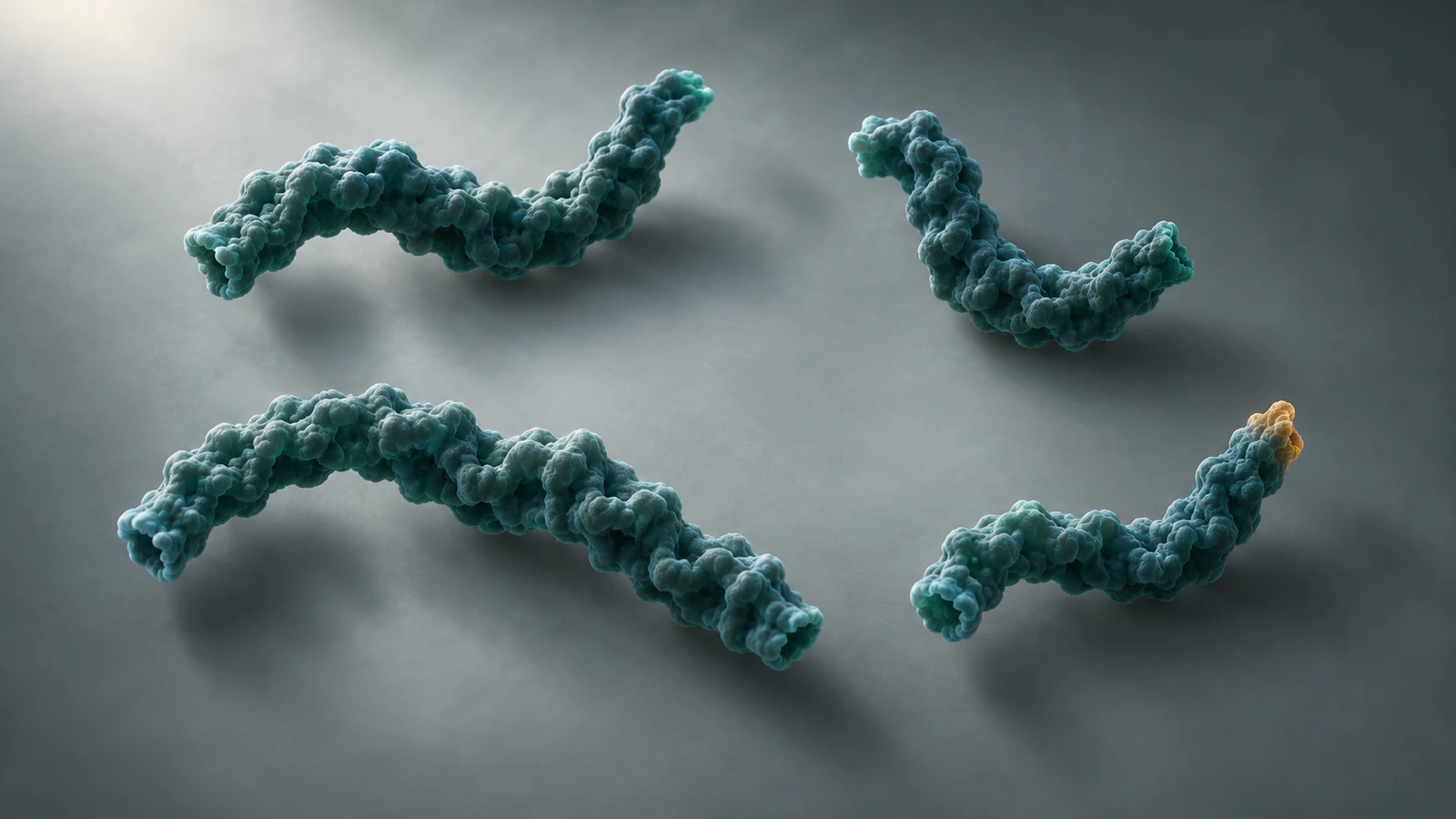
Pause immediately before and after the intended event. In the denaturation branch, trace the backbone between its original ends. In the cleavage branch, identify the cut site and the resulting pieces. If that inspection depends on guessing through a cloud of particles, simplify the shot.
Next review the film without its narration. Can a viewer distinguish one changed chain from several disconnected pieces? Then restore the narration and check that its verbs match the picture: unfolds, associates, cleaves and degrades describe different actions.
Finally check the still images used for a poster or article. Independently generated illustrations may have different geometry and should not be treated as consecutive frames of one measured molecule. Keep their captions explicit and use the animation’s own approved frames when molecular identity across a sequence is important.
- Reject unexplained chain breaks in the unfolding branch.
- Reject new pieces appearing before the illustrated cleavage.
- Reject a cluster that merges independent backbones into one object.
- Reject a glowing effect used as the only evidence of a chemical change.
FAQ
Does protein denaturation break peptide bonds?
Denaturation itself concerns loss or alteration of folded structure while the primary sequence can remain intact. Show backbone cleavage as a separate process, even when harsh conditions can produce multiple kinds of damage.
Is protein unfolding the same as degradation?
No. Unfolding changes conformation; degradation involves cleavage into smaller products. Some degradation pathways include an unfolding step, which is why an animation should distinguish the two operations.
Can a denatured protein refold?
Some proteins can recover under suitable conditions, while others do not. Do not animate automatic recovery for a named protein without evidence that supports the conditions and outcome being shown.
Is aggregation evidence that a protein was cut apart?
No. Aggregation depicts association between molecules. A cluster does not by itself establish peptide-bond cleavage or tell you which chains remain intact.
Can AI-generated protein animation establish a mechanism?
No. It can help communicate a proposed or established explanation, but the scientific claims require independent evidence and review. A plausible-looking chain or enzyme is not a validated molecular structure.
Create a clear unfolding comparison
Start with one intact chain and one reviewable change. Build the denaturation branch in Animiotics, inspect its continuity and then plan the separate cleavage branch with explicitly identified products. Keep the first version focused enough that a colleague can point to the exact frame where its meaning changes.
Try Animiotics with that concrete storyboard in mind. Review the scene against your sources before using it to teach a mechanism or explain a research result.