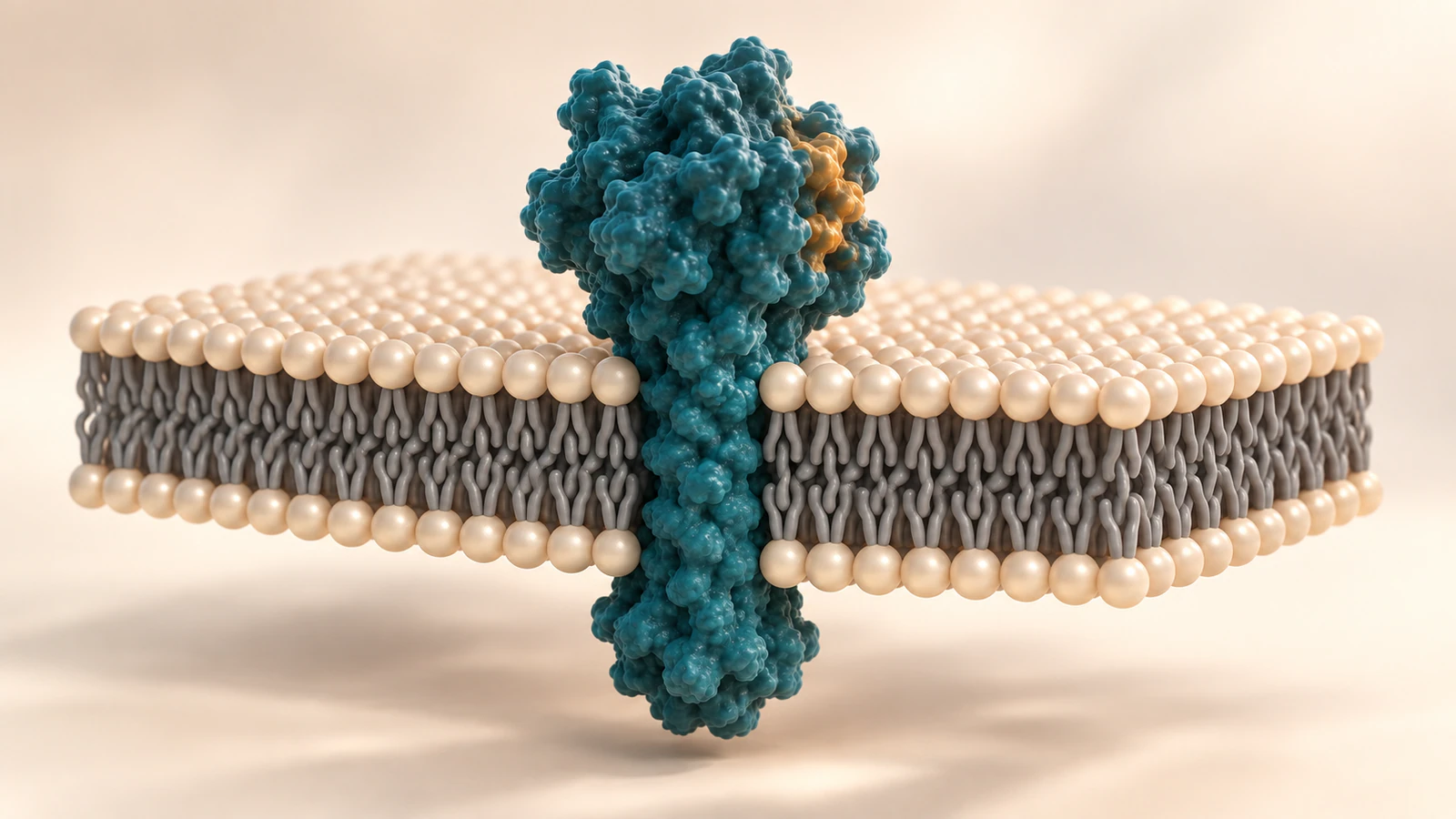
Reveal the interior while keeping the boundary intact
A membrane cutaway animation should make two things clear at once: what the membrane separates and which side each protein domain occupies. Establish those relationships before hiding any surface. Then make the cutaway a visible change in presentation, with no cargo crossing the removed area. Restore the surrounding context before the sequence ends.
This guide is for scientists and science creators planning a membrane-protein scene in Animiotics. Its worked example is deliberately modest: one conceptual protein spans a plasma membrane once, with a larger domain outside the cell and a smaller domain facing the cytosol. The goal is to explain orientation. It does not depict a named receptor, a binding experiment or a transport mechanism.
That limited question gives the animation a useful stopping point. Viewers should leave able to identify both compartments and distinguish an illustration window from a real opening. More molecular detail is valuable only if it helps them make those judgments.
Name the compartments before choosing the camera
The plasma membrane separates a cell from its surroundings and regulates material exchange, as described in the NHGRI plasma membrane glossary. For this scene, call its two sides extracellular space and cytosol. Cytosol means the intracellular fluid outside membrane-enclosed organelles. Avoid using top, bottom, front or back as biological names.
Write the compartment names on the storyboard even if the finished render has no labels. Record where the camera begins and which surface it sees. A camera positioned outside the cell may look down on the protein, but a later camera positioned in the cytosol can look up at the same unchanged object.
For an organelle scene, replace these names with the actual neighboring spaces. Lumen means the space enclosed by the organelle membrane. Calling every visible cavity the cell interior makes it much harder to notice when a soluble object has crossed a boundary accidentally.
Make two leaflets readable at the section edge
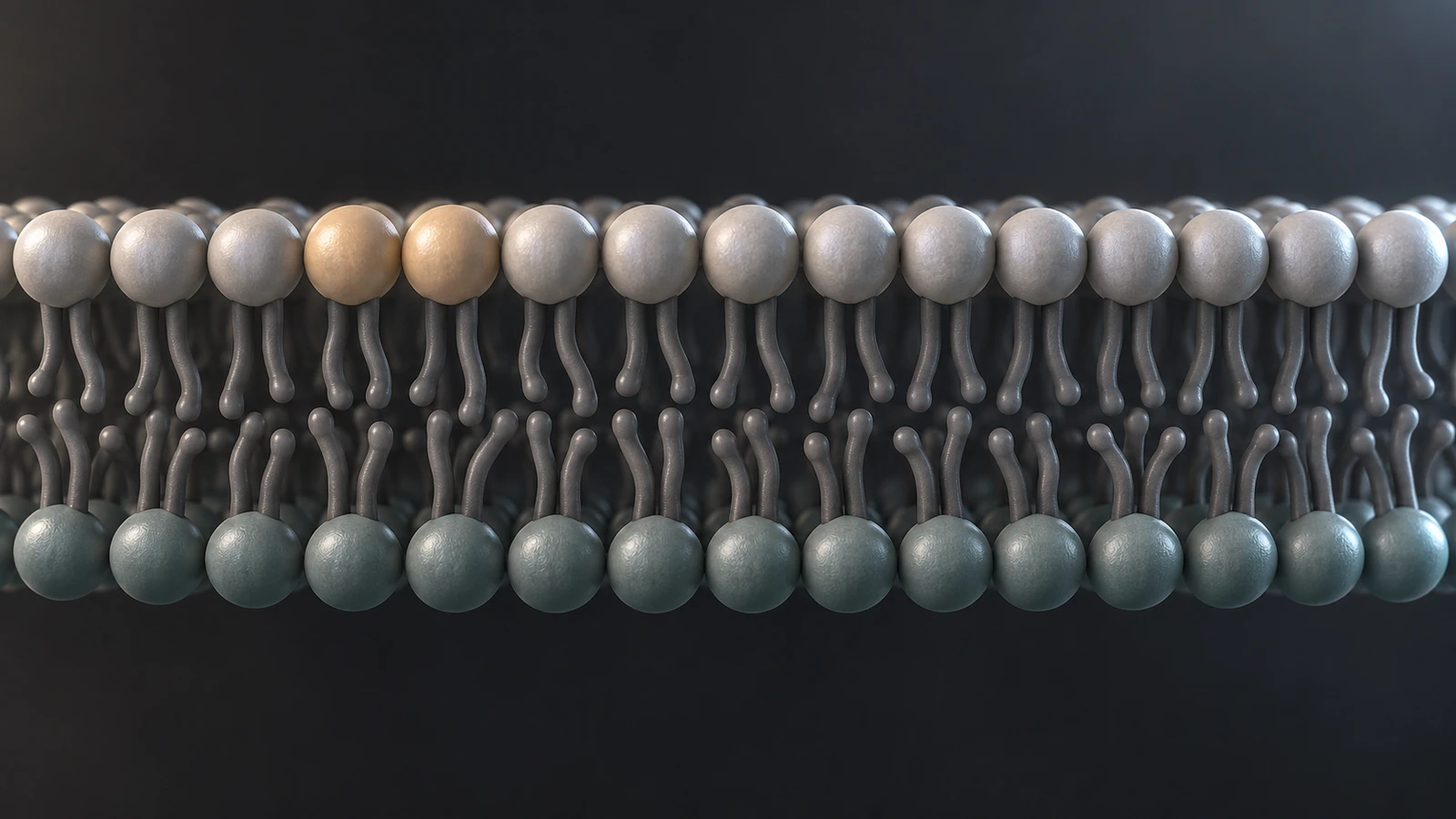
A lipid bilayer contains two opposing leaflets. In a phospholipid teaching model, water-facing heads lie on its two surfaces while hydrophobic tails occupy the interior. The Alberts lipid bilayer chapter explains this organization and why exposed membrane edges are energetically unfavorable. A clean rectangular edge in a render is therefore an illustration convention, not the normal edge of a living plasma membrane.
Inspect the front row of lipids closely. Two opposed sets of short tails should meet within the membrane. Long strands connecting a head on the upper surface directly to a head on the lower surface suggest the wrong molecular construction. Surface polish cannot rescue that mistake.
For a wider shot, simplify the lipids into a thin layered boundary rather than enlarging every head until the membrane dominates the protein. At that distance, the two water-facing surfaces matter more than individual tail details. Explain any exaggerated thickness in the caption and reserve molecular close-ups for a separate view.
Check orientation evidence for a named protein
The example here assigns orientation as part of a conceptual scene. A named protein needs a different starting point: identify its species, sequence or isoform, membrane location and available topology evidence before arranging its domains. A visually attractive model does not establish which end faces which compartment.
UniProt topological-domain documentation describes annotations for the non-membrane regions of membrane-spanning proteins. It also distinguishes experimental and predicted information. Even when a compartment assignment has experimental support, the precise boundaries of an annotated region may be predicted. Record the evidence attached to the feature you actually use.
Make a short asset note with three entries: supported orientation, uncertain boundaries and deliberately simplified geometry. If only an isolated extracellular domain is available, do not stretch it through the membrane to manufacture a complete receptor. Show the supported fragment and explain the missing context, or use an explicitly conceptual whole-protein illustration.
Choose a cutaway that cannot be mistaken for a pore
A cutaway removes part of the displayed surface so the viewer can see behind it. A pore is a physical passage in the biological subject. Those two ideas require different visual treatment. For an orientation explainer, place the editorial section at the front boundary of the scene, away from any feature that could be read as a transport opening.
Begin with an intact overview, pause the biological action and reveal the section. Keep the protein and nearby objects stationary while the display changes. This isolates the reason for the new view. If the scene begins with a glowing circular hole around the protein, viewers may reasonably interpret it as an opening the protein created.
Transparency can help with a shallow obstruction, but several transparent surfaces may become harder to interpret than one clean cutaway. Choose the smallest display change that reveals the relevant relationship. For a related representation decision, see protein surface versus ribbon.
Use a compartment ledger for every moving object
Before animating, assign each object a location and a rule for whether that location may change. This is a production checklist, not a biological database. Its purpose is to expose contradictions while they are still cheap to fix. The orientation example needs only four rows.
Write the extracellular domain as outside throughout; write the cytosolic domain as cytosolic throughout; write the membrane-spanning segment as embedded throughout. Finally, list the camera separately with permission to change viewpoint. The camera is not a molecule and does not need a biological route through the membrane.
Add any later cargo as a new row rather than treating it as background decoration. State its starting compartment, ending compartment and the source supporting a crossing event. If no crossing is part of the question, keep it on its assigned side. A particle drifting through the editorial section can accidentally become the most consequential claim in the entire film.
Storyboard a five-shot orientation explainer
Use the following sequence as a concrete first pass. Its durations are editorial suggestions, not measured biological times. Adjust them after viewing the result at presentation size. The central discipline is to change one explanatory variable at a time.
Shot one, about four seconds: establish the membrane patch and the whole protein in an oblique view. Use the larger outer domain as the recognizable landmark. The viewer should see a continuous boundary before any section is revealed.
Shot two, about three seconds: freeze the subject and reveal the front section. Keep lighting and camera position stable. State in narration or a caption that the membrane is cut away for visibility. No particle should move through the newly visible region.
Shot three, about five seconds: move closer to the embedded segment. Preserve the relationship between both domains and the two membrane faces. Use a restrained accent to guide attention, without presenting the colored patch as a measured binding site.
Shot four, about four seconds: change the viewing angle enough to show the smaller cytosolic domain. Keep the same protein geometry and orientation relative to the membrane. The camera movement should reveal an existing feature rather than make the protein appear to invert.
Shot five, about four seconds: return to the overview and restore the intact display. End on the same compartment arrangement established in shot one. The final frame should make sense to someone who sees it alone, without relying on the missing section as the only orientation cue.
Preserve sidedness when extending the story to vesicles
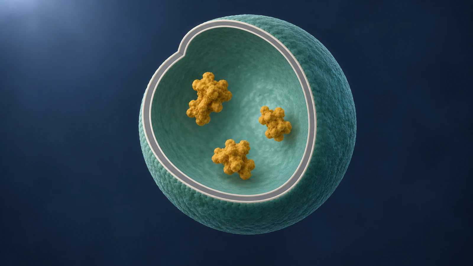
A vesicle scene introduces a distinction that a flat membrane patch can hide: being geometrically inside the cell does not make a compartment cytosolic. In the secretory pathway, membrane-protein orientation is retained during vesicular transport. Domains facing the ER lumen correspond to domains facing the extracellular space after delivery to the plasma membrane. Cooper’s ER chapter describes this continuity.
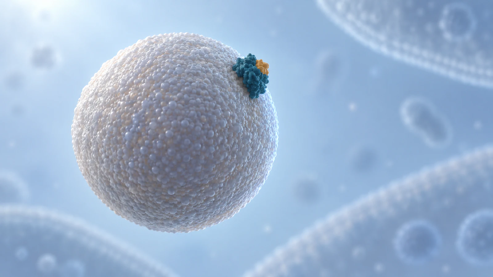
If you extend the storyboard, track the labeled domain through each change of membrane shape. Do not rotate the protein independently simply to keep its highlighted part facing the viewer. Adjust the camera instead. Use a cutaway of a vesicle only after a complete-shell view has established that its lumen is enclosed.
Keep this extension separate from a detailed budding or fusion mechanism. A shape transition between two illustration states is insufficient evidence for the molecular steps connecting them. For an orientation lesson, a clearly explained pair of endpoint views may be more honest and more readable than an elaborate invented transition.
Keep scale, material and motion subordinate to the question
Use a restrained palette with one stable accent on the domain the viewer needs to follow. Do not switch that accent between compartments when the camera moves. Color is an editorial guide here, not a claim about natural molecular appearance or biochemical activity.
Choose enough surface texture to separate protein from membrane without making every lipid a competing hero. A soft key light and controlled edge light can reveal the smaller cytosolic domain without turning the entire membrane into glass. Check the darkest view separately: losing one membrane face in shadow can destroy the orientation lesson.
Keep apparent size changes attributable to the camera. If a local protein view requires a different scale from the opening, make that transition explicit. The molecular scale guide provides a complementary way to separate object dimensions, screen size and playback time.
Review the boundary before approving the render
Scrub the draft with the sound muted. At the intact opening, the maximum cutaway and the final restored view, ask a colleague to identify the extracellular and cytosolic sides. Ask whether anything crossed the membrane. If their answer differs from the storyboard, fix the image before adding more narration.
Then inspect still frames at full resolution. Check the bilayer midplane, the continuity of the embedded protein and the edge of the editorial section. Look for unintended holes, detached domains or cargo touching a boundary it should not cross. Repeat the check at thumbnail size to see whether the larger spatial relationships survive.
Keep a short approval note: orientation checked against the asset record; display removal distinguished from biology; no unsupported crossing; scale changes explained. Captions should identify conceptual geometry and explain the removed surface. These checks make the review concrete without pretending that a polished illustration is experimental evidence.
FAQ: membrane cutaways and protein orientation
Does a membrane cutaway depict damage?
It should not unless damage is the subject. Establish an intact boundary first and identify the missing surface as an illustration choice. Keep objects from crossing the cutaway so the visual does not imply leakage.
Should the extracellular side always be at the top?
No. Top is a screen position, while extracellular names a biological compartment. Choose a clear initial view and preserve orientation as the camera moves. Keep compartment names in the storyboard even when labels are absent from the render.
Can a conceptual protein be used for this lesson?
Yes, if it is clearly described as conceptual and the assigned orientation is stated. Do not attach a named receptor, structural identifier or binding claim to an illustration that was not built and checked for that identity.
Is a vesicle lumen the same as cytosol?
No. Treat an enclosed lumen as a separate compartment in the storyboard. For a secretory-pathway orientation lesson, follow the membrane-facing relationships through the endpoint views rather than renaming every space inside the cell as cytosol.
How much lipid detail should be visible?
Enough to support the current question. Show two opposed leaflets in the close section view, then simplify the boundary in a wide shot. The viewer should recognize the membrane and both protein domains without counting individual lipids.
Try a focused protein animation in Animiotics
Use Animiotics to create a short protein overview for your next membrane explanation. Start with one supported protein model and decide which domain the audience should recognize first. Use the five-shot plan as a review brief, then check the resulting views against your compartment ledger. Treat any membrane cutaway as an editorial construction that needs its own verification.
Signing up and the manual editor are free, and new customers can try AI generation for $2.99. Begin with one clear orientation question and a small scene you can review carefully before extending it into a mechanism.