Start with the claim your animation must explain
An enzyme inhibition animation should tell viewers where an inhibitor binds, which molecular states your model permits and what the experiment measures. Decide those three things separately before building the scene in Animiotics. A striking pocket close-up can explain location while leaving the kinetic mechanism unresolved.
The most useful comparison is often smaller than a complete drug story: a substrate approaching an occupied site versus a substrate remaining bound while an inhibitor occupies a separate site. These are proposed visual situations, not sufficient evidence for assigning a kinetic class. The storyboard below helps you show them without turning an illustration into a stronger claim than your sources support.
Use this guide when briefing a short explainer for a research presentation, teaching session or scientific product page. Its deliverable is a scene contract that a scientist and an animator can both review. You should leave with clear decisions about occupancy, camera continuity and the evidence that belongs beside the finished animation.
Keep location and kinetic behavior in separate sentences
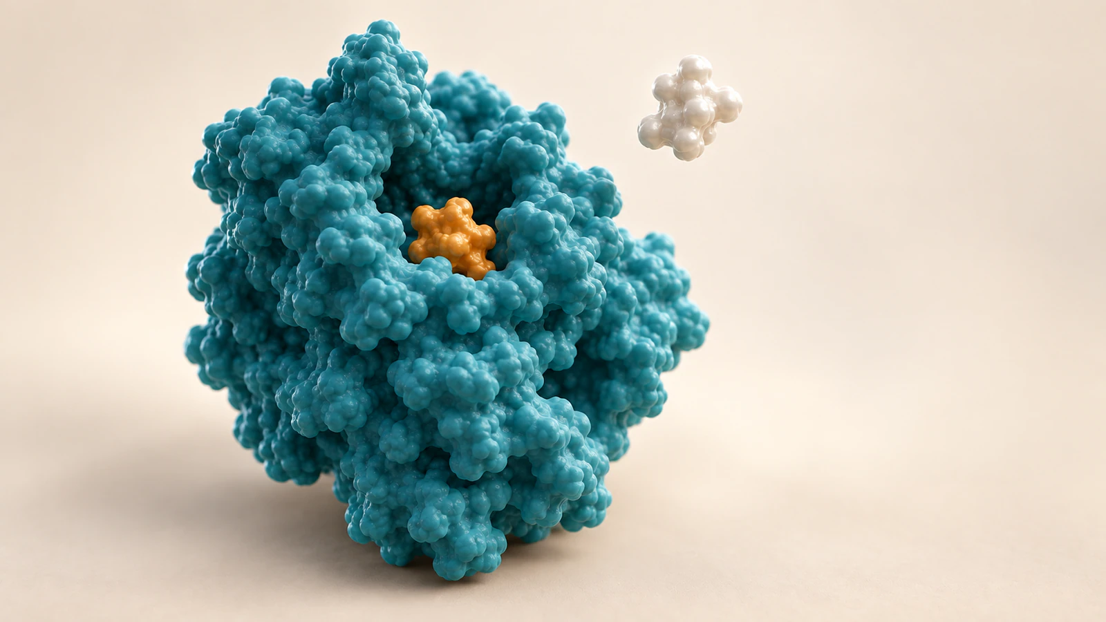
A substrate is a molecule on which an enzyme acts; a ligand is any molecule that binds to a target. Competitive inhibition means inhibitor and substrate binding are mutually exclusive in the model. Sharing a pocket is a familiar example, not the universal definition. Allosteric inhibitors act through regulatory interactions and can produce different kinetic patterns. The Assay Guidance Manual explicitly separates these ideas.
Write two sentences in your brief: “The inhibitor occupies this supported location” and “The assay supports this behavior with respect to this substrate.” If the first sentence is unknown, do not invent a precise pocket. If the second is unknown, explain binding without assigning a kinetic label. A remote amber object is a location cue, not a result.
For a general teaching scene, introduce the protein and its active site, the region involved in catalysis, before introducing the inhibitor. Let the viewer establish orientation. Reserve one color for the inhibitor throughout the sequence and give the substrate a different silhouette as well as a different color.
Use pure noncompetitive carefully
In the ideal pure noncompetitive model, an inhibitor binds equally well to free enzyme and the enzyme–substrate complex. Apparent Vmax decreases while apparent Km stays unchanged. Those conditions are more specific than “binds somewhere else.” They should come from an appropriate kinetic analysis, not from simultaneous occupancy in a frame. Assay Guidance Manual.
Vmax is the limiting reaction rate; for Michaelis–Menten kinetics, Km is the substrate concentration at half that rate. Km is not generally a binding dissociation constant. The IUBMB terminology recommendations also warn that “noncompetitive” has been used ambiguously for mixed inhibition. Name the intended model explicitly.
This vocabulary affects the edit. Do not title a sequence “noncompetitive” merely because a substrate is visible beside a bound inhibitor. Use “illustrative remote-site binding” while the mechanism remains unassigned. That working title gives the scientific reviewer a specific question to resolve before voiceover or captions make a definitive claim.
Build a site, state and evidence contract
Before choosing camera moves, fill in one row for each proposed scene. The contract below is an editorial tool, not an assay protocol. It forces the team to distinguish what appears on screen from what justifies the explanation. An empty evidence cell means the scene needs a narrower claim or a visible qualification.
Keep this document beside the storyboard. During review, ask for corrections to a particular row rather than general approval of “the science.” A reviewer might accept the binding site but reject the proposed motion, or accept a conceptual state while asking you to remove an unsupported efficacy statement.
| Contract field | Record before animation | Decision it controls |
|---|---|---|
| Site | Supported pocket, surface region or explicitly unknown location | Whether a close-up can name or highlight a particular site |
| State | Which objects may be bound together in this selected model | Whether substrate and inhibitor may share the same frame on one enzyme |
| Evidence | Structure, assay or hypothesis supporting each claim | What the caption may conclude and what stays illustrative |
| Continuity | Stable protein view, ligand identity and chosen orientation | Whether viewers can track the comparison across cuts |
| Limits | Unmeasured geometry, timing and particle counts | Which details must not look quantitative |
Work through a five-shot comparison
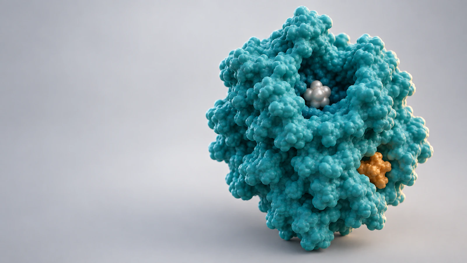
Imagine a teaching brief comparing a selected same-pocket competitive model with a selected remote-site model that permits simultaneous binding. It does not name a drug or claim that the second model is pure noncompetitive. Use one generic teal enzyme envelope, pearl substrate and amber inhibitor. Treat both models as conceptual until you replace their assumptions with evidence for a real system.
Shot one establishes the protein in a three-quarter view. Hold the camera long enough to locate the cleft and identify the substrate. Avoid an orbit during this introduction: a continuously turning protein makes it harder to remember which depression mattered. Use a restrained change in framing to make the first close-up feel connected to the opening.
Shot two introduces the same-pocket example. The inhibitor occupies the selected site while the substrate stays outside. Keep a clear visible gap rather than allowing the substrate to overlap the protein for dramatic effect. Do not show the two ligands wrestling or one molecule physically pushing the other away. The teaching point is exclusion in the chosen model.
Shot three resets to the original orientation before switching examples. Make the reset explicit with narration or an editorial transition. Without it, a viewer may interpret the inhibitor moving across the protein as a measured migration pathway. This is a comparison of models, so the transition should not masquerade as a continuous molecular event.
Shot four shows the remote-site example with both ligands visible in their separate locations. Keep any proposed conformational change modest and identified as schematic unless supported by the selected structural evidence. A camera move can reveal a hidden ligand; it should not create the impression that the site appeared only after binding.
Shot five moves to the evidence statement. For a real project, use the approved experimental result with its conditions and source. For this conceptual exercise, end by stating which binding states were illustrated and which kinetic questions remain open. Do not add an invented percentage, activity curve or stream of products just to give the animation a decisive ending.
Choose structures without borrowing the wrong biology
A real structural example can sharpen the brief even when it is not your animation subject. The RCSB PDB-101 account of aspartate transcarbamoylase describes E. coli ATCase with catalytic and regulatory chains, and CTP binding that stabilizes its T state. It also explains that human ATCase has a different organization. Species and assembly therefore belong in the asset record.
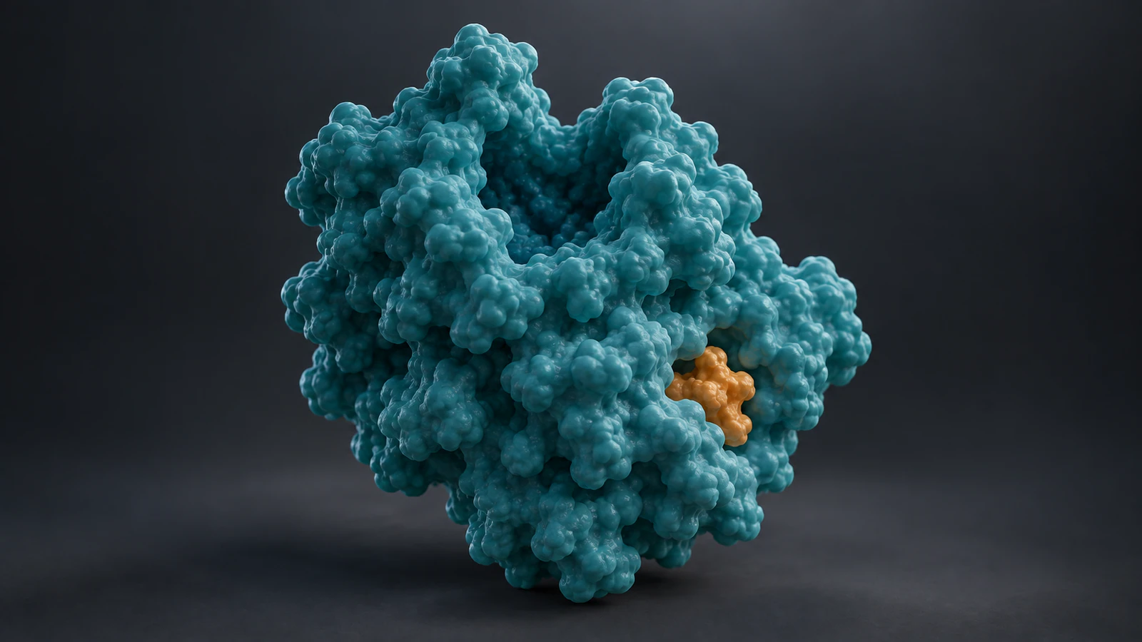
PDB entry 4FYW is E. coli aspartate transcarbamoylase complexed with CTP. It is a specific reference to inspect, not the identity of the generic enzyme pictured here. These images were created as conceptual illustrations; they were not rendered from that entry’s coordinates.
For your own structure, record organism, construct, assembly and bound ligand before requesting an accurate molecular asset. Keep that record attached when the scene changes hands. A beautifully finished generic surface should never acquire a PDB identifier simply because its overall silhouette resembles the named protein.
Direct the camera around the binding question
Frame the cleft so the occupied region remains legible at presentation size. If the inhibitor disappears behind the surface, first try changing the viewing angle. A transparent shell may solve visibility while introducing ambiguous overlaps. Our guide to protein surface versus ribbon views can help you choose the representation for that particular question.
Keep the inhibitor’s material and silhouette consistent across the cover, close-up and final state. A small ligand should not become protein-sized simply to dominate the frame. Enlarge the camera view instead. When the exact dimensions are unknown, avoid a scale bar and describe the view as conceptual.
Use lighting to separate the molecules without assigning it a biological meaning. An amber inhibitor against a teal protein can be easy to follow on a dark background. A pearl substrate may need a darker local surface or a slightly different angle on a light background. Test each frame as a thumbnail before approving fine surface detail.
Show an ensemble without inventing concentration
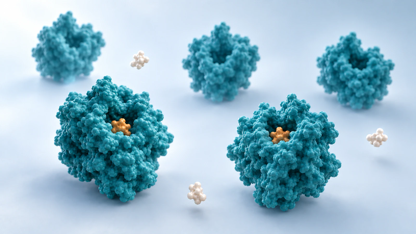
A wider view with several enzymes can support a transition from an individual binding event to a population-level question. Keep the foreground sparse enough that the audience can identify different occupancy states. Do not arrange molecules into a neat queue that suggests they deliberately wait for a turn at the enzyme.
Write down what the particle count means. In a conceptual scene it is a composition choice, so the caption should say it does not represent concentration or occupancy measurements. If a project requires quantitative populations, those numbers need their own data and mapping rules before animation begins.
For more help separating visual timing from physical quantities, read molecular scale in animation. In this storyboard, a longer hold gives the viewer time to inspect a pocket. It does not establish a residence time, reaction rate or molecular trajectory.
Review the scene before polishing the finish
Run one scientific review with the site/state/evidence contract visible, then a communication review with the sound muted. The first checks the claim. The second checks whether viewers can identify the same molecules across cuts without relying on narration to repair the picture.
Freeze the opening, occupied-site shot and final bound-state view. Ask a reviewer to point to the substrate and inhibitor in each. If they need to guess, fix the orientation, contrast or object identity before spending time on atmosphere. Compare the final caption with the contract as well: a cautious storyboard can still be undermined by an overstated caption.
- Does every named binding location have evidence or an explicit conceptual label?
- Does the selected model permit every simultaneous occupancy shown?
- Does the edit distinguish a comparison reset from a continuous pathway?
- Are the scientific result and the visual interpretation clearly separated?
- Would the scene still communicate its intended question as a small still image?
FAQ: planning an enzyme inhibition animation
Does allosteric always mean noncompetitive?
No. Keep the binding explanation and the kinetic classification separate in the brief; a remote-site image alone does not assign the class.
Can I use a generic enzyme shape?
Yes, for an explicitly conceptual explanation. Do not label it as a named structure or use its invented pocket geometry as evidence for a real binding mode.
Should substrate and inhibitor appear bound together?
Only when the selected model permits that state. Write the allowed occupancy into the storyboard contract before asking for the frame.
How should I handle an unknown binding site?
Keep the location unresolved in the visual and caption. You can still explain the measured finding without inventing a precise pocket or a molecular pathway.
Do I need a kinetic plot in the animation?
Only if it answers the intended question and you have a suitable sourced result. A conceptual comparison can end with an explicit statement of what remains unmeasured.
Try a focused binding comparison in Animiotics
Start with a short enzyme animation in Animiotics: establish the protein, reveal an occupied site and finish with one clearly explained binding state. Use the contract above as your scientific brief and review the resulting visuals against it. Keep the first project narrow enough that a viewer can follow the substrate and inhibitor from beginning to end.