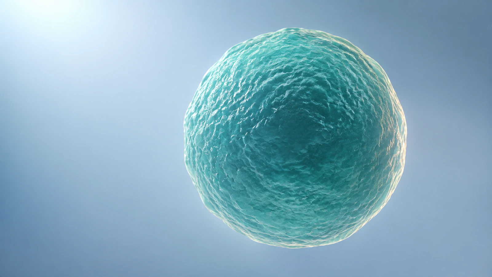
Make scale a decision before you animate
Molecular scale in animation becomes clear when each shot has a defined field of view and each transition tells the viewer that the viewing scale has changed. Keep relative dimensions consistent within a shot. Use a cut or a clearly explained magnification when moving from a cell to a protein. Treat playback duration as a separate editorial choice unless you have evidence that supports a biological clock.
This matters because the most readable composition can also be misleading. A large antibody beside a fully visible cell may look attractive, but the shared space suggests comparable dimensions. A smooth five-second approach to a receptor can look like a measured binding trajectory. Neither implication is justified simply because the objects are recognizable.
The workflow below uses a hypothetical extracellular binding scene. It provides a scale contract, a numerical framing example, and a five-shot storyboard you can adapt before building the final animation. The accompanying CGI images are conceptual illustrations, not calibrated scientific images or coordinate-derived molecular structures.
Separate physical size, screen size, and resolution
Physical size describes the object in units of length. Screen size describes how many pixels it occupies in the final image. Resolution describes what detail the underlying evidence or imaging method can distinguish. These quantities interact, but they are not interchangeable. Enlarging a model on screen does not add structural evidence.
The SI prefixes micro and nano represent one millionth and one billionth, respectively. Therefore, one micrometer equals one thousand nanometers. These definitions come from the BIPM SI prefix table. Convert all source dimensions into one unit before comparing assets; otherwise, a correct number attached to the wrong unit can produce a thousandfold error.
For biological context, the opening of this cell-biology chapter describes a typical animal cell as 10–20 micrometers across. That is a useful order-of-magnitude reference, not a dimension to impose on every cell. Cell type, state, and the particular specimen still matter. Record the actual source when your scene represents a named system.
Use one calculation to test the opening composition
Choose a deliberately simplified example: a cell 10 micrometers across and a molecular envelope 10 nanometers across. The molecular dimension is a teaching assumption, not a measurement of the antibody illustrated here. Expressed in nanometers, the cell spans 10,000 nanometers. Its diameter is one thousand times the chosen molecular dimension.
If that cell occupies 1,000 pixels across the frame, the molecule occupies roughly one pixel when both lie at the same depth and use the same linear scale. A recognizable, 100-pixel molecular hero beside this cell would therefore require a different framing scale or an explicit illustrative enlargement. It cannot silently remain an equal-depth, same-scale view.
For a separate close-up spanning 100 nanometers across 1,000 pixels, the same 10-nanometer envelope occupies about 100 pixels. This is the useful production decision: change the field of view instead of secretly enlarging only the molecule. These numbers are calculated framing examples; perspective, cropping, and a different output width will change the screen measurements.
| View | Assumed field across 1,000 pixels | 10 nm envelope on screen | Editorial use |
|---|---|---|---|
| Whole cell | 10 micrometers | About 1 pixel | Locate the biological setting |
| Local molecular view | 100 nanometers | About 100 pixels | Explain a molecular relationship |
Write a scale contract for every shot
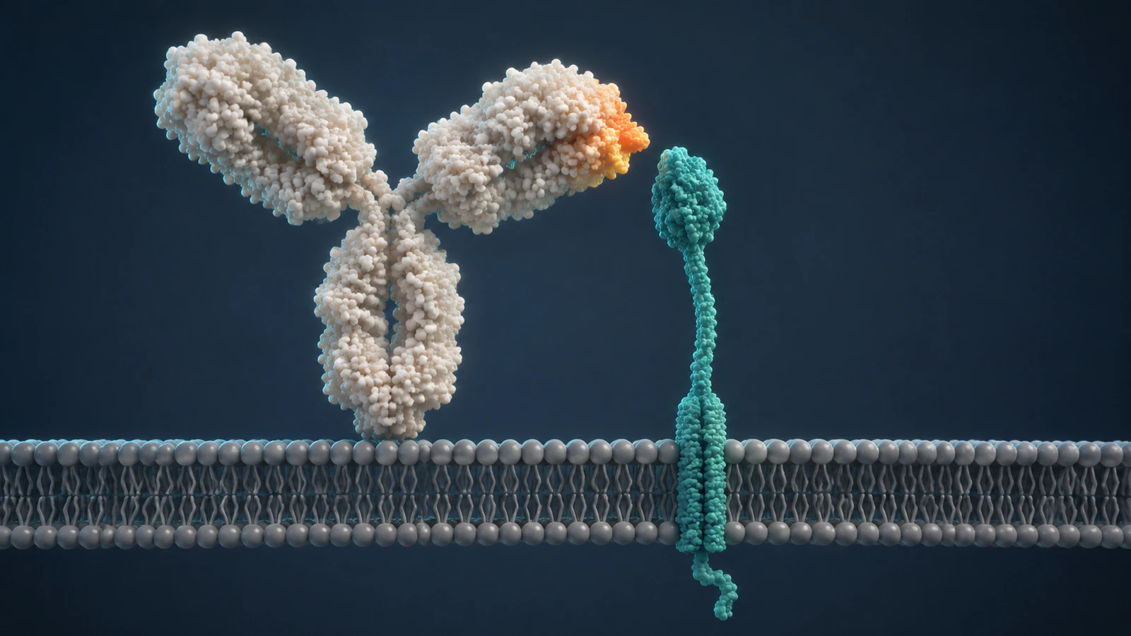
A scale contract is a short production note that makes the assumptions inspectable. It travels with the storyboard, not just with the final render. Begin with the biological compartment and the camera field of view. Then identify one reference object whose dimension you can trace, along with the source and any uncertainty.
Record whether the shot is calibrated, approximately proportional, or intentionally enlarged for explanation. A calibrated claim needs a defensible mapping between model coordinates and physical units. An approximate scene can preserve useful relationships without implying a measurement. An enlarged explanatory view needs a disclosure wherever its exaggeration affects interpretation.
Finish the note with what changes at the next cut. The camera may magnify a membrane region while the model proportions stay fixed. Alternatively, the sequence may switch to a separate schematic scene. Writing that distinction now prevents the animator, reviewer, and caption writer from solving three different versions of the same shot.
- Compartment and purpose: where the action occurs and the one relationship the viewer should understand.
- Reference and units: source object, physical dimension, assembly choice, and conversion convention.
- Representation: surface or ribbon, omitted structures, and whether proportions are calibrated or illustrative.
- Transition and time: what changes at the cut, and whether playback has any measured time meaning.
Check molecular identity before calibrating the asset
A familiar silhouette is not enough to establish molecular identity. For an experimental model, inspect the organism, construct, chains, biological assembly, and experimental method. A file may contain only part of the object your narration names. Calibrating that fragment perfectly will not make it an intact molecule.
For example, RCSB PDB entry 1IGT describes an intact mouse IgG2a antibody, Mab231, determined by X-ray diffraction at 2.80 angstrom resolution. Its listed biological assembly is an A2B2 heterotetramer. It is a specific structural record, not a generic human therapeutic antibody or a receptor-bound complex.
The resolution value is not the overall antibody width. Nor does the structure establish how long binding takes. If you use this entry, document the assembly and measure the relevant envelope from the coordinates using an appropriate structural viewer. Do not copy a width from this article’s hypothetical example. Our CGI antibody-like shapes are not reconstructions of 1IGT.
For preparation decisions before this calibration step, see PDB to animation. For choosing which parts of an imported structure remain visible, use the separate surface versus ribbon guide.
Build a five-shot sequence with visible changes of scale
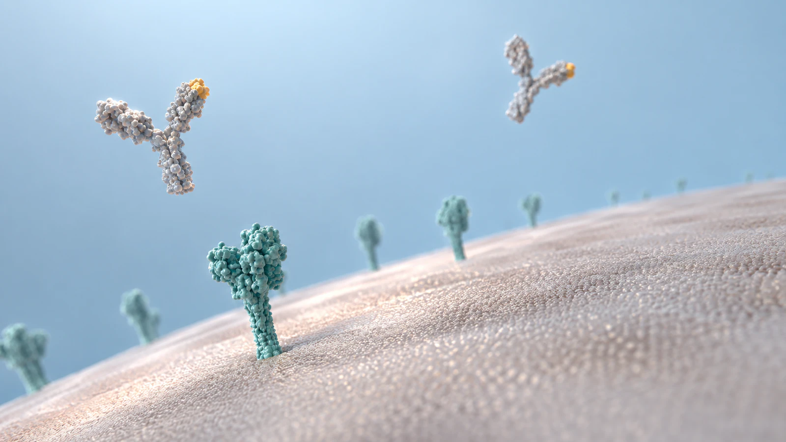
Start with the whole cell to answer where. Then cut to a local membrane view to answer which compartment. Only after that orientation is stable should the binding partners become the hero. A cut is often easier to interpret than an uninterrupted camera flight that changes scale by orders of magnitude.
The storyboard below is an original production framework for a hypothetical interaction. It does not assert a particular antibody–receptor pairing, binding pose, or kinetic mechanism. Replace the generic objects with evidence-supported assets when a real system is being explained. Keep the local membrane orientation recognizable between adjacent shots.
| Shot | Visible content | Scale and evidence note |
|---|---|---|
| 1. Locate | One complete cell with an identifiable membrane region | Cellular context; no enlarged individual antibodies beside the whole cell |
| 2. Reorient | A local membrane patch and the extracellular space | Explicit magnification; preserve which side faces outside |
| 3. Identify | The two molecular partners before contact | Use one local coordinate scale; name any conceptual geometry |
| 4. Explain | A close view of the relationship being discussed | Explanatory event unless a supported trajectory is available; no invented clock |
| 5. Return | A wider local view with the interaction still situated | Restore context without implying a measured population response |
Keep camera changes from becoming biological changes
Perspective makes nearby objects look larger. If the hero molecule moves toward the camera while the receptor remains farther away, their apparent size ratio changes even when their model dimensions do not. That can be useful cinematic depth, but it is a poor view for judging physical size. Use a common depth plane for a comparison shot.
Avoid moving the camera, changing focal length, rescaling the hero, and rotating the membrane simultaneously. Reviewers then cannot tell whether the molecule changed size, the camera moved, or the scene switched to a different representation. Make the transition legible by holding one recognizable spatial reference stable.
A scale bar also needs a defined interpretation. In a perspective scene, a bar calibrated at one depth does not describe every object in the volume. Tie it to the relevant plane or use a separately calibrated comparison view. If the geometry is conceptual and unmeasured, a numerical bar adds false precision; a clear caption about the change of view is more honest.
Give playback time its own evidence rule
A frame rate tells you how images are played, not how rapidly the biology proceeds. A structure file contributes spatial information; it does not supply a binding clock. If you choose a slow approach so viewers can recognize the partners, describe that segment as explanatory timing rather than assigning it a real duration.
When working from an actual trajectory or time series, keep the sampling interval, selected frames, playback rate, and any pauses in the production record. A hold added for narration breaks a simple constant relationship between screen time and source time. A numerical time annotation must account for that choice.
For an illustrative sequence, avoid precise rate claims altogether. State the order or relationship that the evidence supports and disclose that timing is simplified. If the evidence establishes only a before-and-after state, the interpolated movement between them remains an animation choice. A convincing transition cannot supply the missing experimental observation.
Review the sequence at delivery size
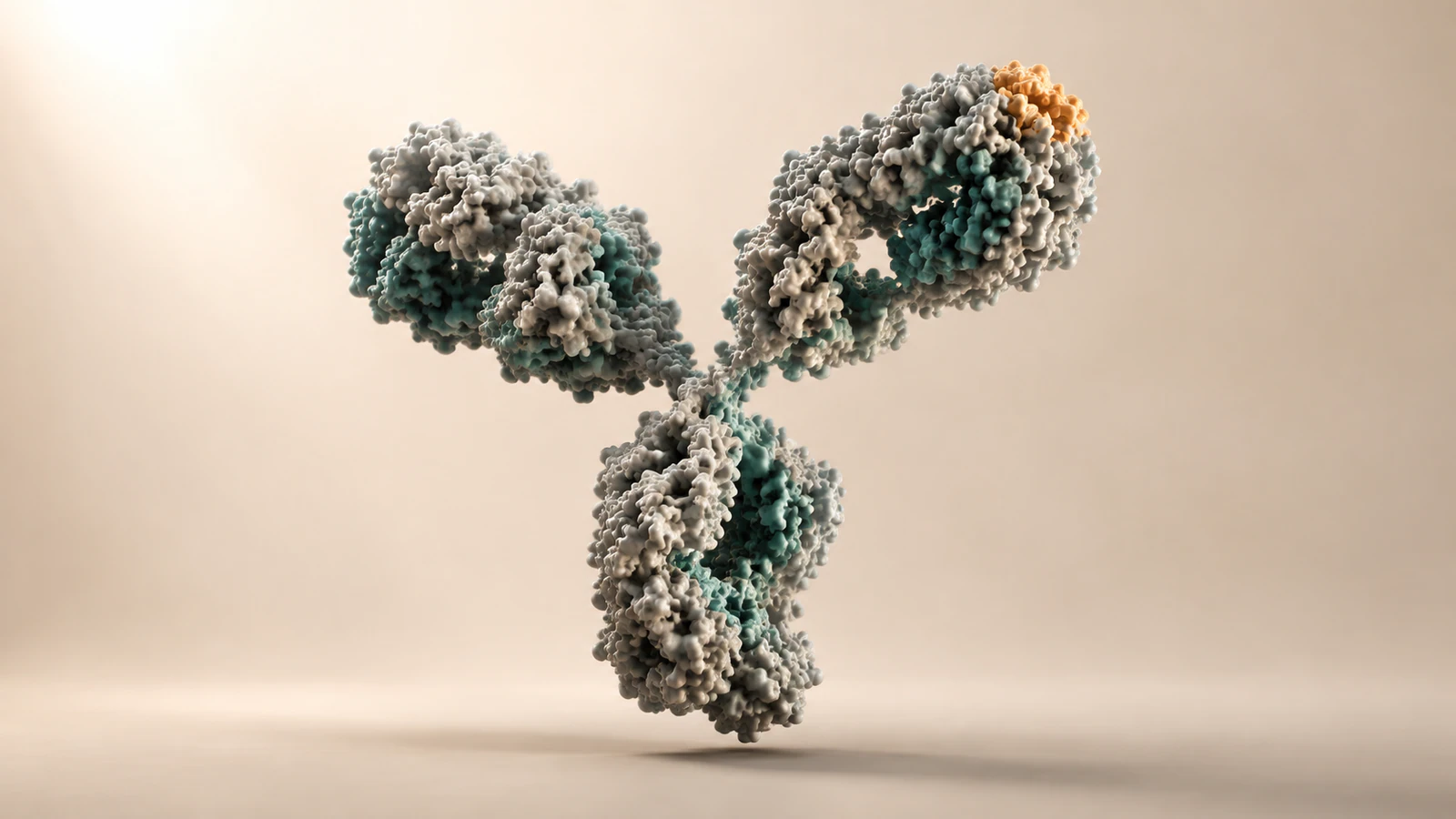
Review a small draft before spending time on final materials. First mute the narration and ask a colleague to describe the size relationships. If they infer that the antibody is a substantial fraction of the cell, the visual explanation failed even if a later voiceover gives the right numbers.
Next inspect the transition frames individually. The extracellular side should remain identifiable; a cropped membrane edge should not look like a biological hole; and a local interaction should not silently become a whole-cell outcome. Check that any contact shown belongs to the stated evidence level.
Finally, read the captions with the images hidden. They should identify what is conceptual, what is calibrated, and which numerical examples are assumptions. The four illustrations here teach framing choices; none can serve as a molecular ruler. Keep the scale contract and source record with the project so a later crop or reuse does not strip away that context.
FAQ: molecular scale and animation timing
Must every scientific animation be exactly to scale?
No. Explanatory scenes can simplify or enlarge objects, provided the important relationships remain defensible and the change is disclosed. Decide which dimensions affect the reader’s conclusion before choosing the level of calibration.
Can I show a whole cell and a detailed protein together?
Yes, as clearly separated views or an explicitly enlarged illustration. In one shared spatial view, check their physical ratio first. A protein large enough to recognize may imply the wrong size if the cell is also fully visible.
Does a PDB resolution value tell me the molecule’s diameter?
No. Experimental resolution and the overall spatial extent of a model describe different things. Check the correct assembly and measure the relevant model dimension instead of treating resolution as object size.
Does smooth movement count as a molecular simulation?
No. Keyframed motion can explain an idea, but smoothness does not establish a physical model, a sampled trajectory, or measured kinetics. Identify the source of the motion and label illustrative interpolation accordingly.
What should I do when the source gives no timing?
Use explanatory pacing and say that the timing is simplified. Do not attach seconds or milliseconds to the biology merely because the finished clip has that playback duration.
Try a cell-to-molecule scene in Animiotics
Use this storyboard to plan one short cell-to-molecule animation in Animiotics: establish the cell, reorient at the membrane, and explain one local molecular relationship. Write the scale contract first, then check the draft at its intended viewing size. Keep the numerical teaching example separate from any dimensions you measure for your own assets.