Find the gap before drawing the loop
To handle ChimeraX missing residues, compare the deposited sequence with the residues that have coordinates, then inspect the missing-structure pseudobonds. These connectors mark an absent segment; they do not recover its atomic positions. For a research figure, leave the interval open or use a clearly explained dashed connector. Record the missing residue numbers in the caption before making an animation.
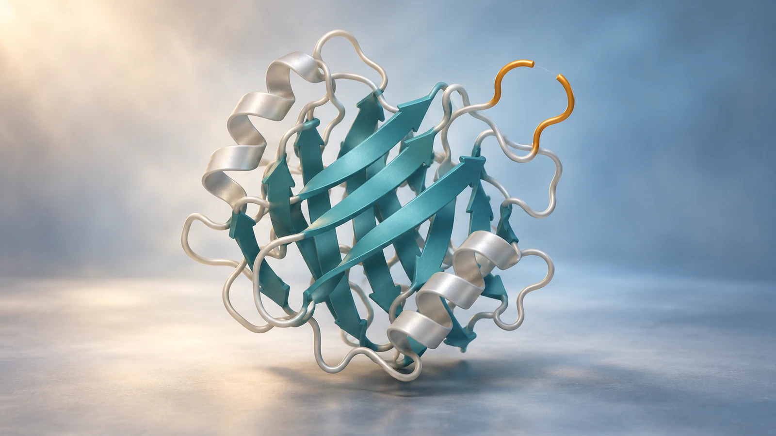
This guide uses the real PDB entry 1FAT. Its chain A has an internal omission at Gly37 and a missing terminal segment, giving you two different cases to check. You will produce a short gap audit and a reproducible display setup. The illustrations are conceptual, not renders of 1FAT or evidence for the shape of its missing regions.
Prepare a traceable starting file
Download the PDBx/mmCIF file from the 1FAT entry and keep an unchanged copy. PDBx/mmCIF stores coordinates and descriptive tables in a text file. Note the accession, download date, chain identifier and software version in a small companion record. Use a fresh ChimeraX session so the first structure receives model number #1.
The official news page lists ChimeraX 1.12 as a production release. We checked current documentation and executed the coordinate audit, display commands and session save in installed ChimeraX 1.9 without its graphical interface. Interactive framing, Sequence Viewer appearance and image export below are documented steps, not locally inspected renders.
1FAT is phytohemagglutinin-L from common bean, determined by X-ray diffraction at 2.80 angstrom resolution. We use one chain to demonstrate coordinate completeness, not to represent the full biological assembly. For assembly choices, use the separate protein-complex guide.
Separate three kinds of absence
A blank region in a picture can have several causes. The atoms may exist in the loaded model but be hidden. A sequence position may be represented in the experimental construct yet lack coordinates. Alternatively, a domain may never have been included in the construct used for structure determination. Those cases require different explanations.
The PDB-101 missing-coordinates guide explains why flexible regions can be unresolved and why some entries contain only fragments. Absence alone does not establish a particular motion, cleavage event or folding state. Check the entry and associated study before assigning a biological cause.
Make the distinction explicit in the working record before changing the picture. A presentation choice can hide an atom; it cannot supply a missing experimental coordinate.
| What you find | What to check | Figure treatment |
|---|---|---|
| Present coordinates, hidden display | Atom and cartoon visibility | Restore the relevant display |
| Unobserved sequence positions | Entry annotations and chain sequence | Open interval or explained connector |
| Sequence outside the construct | Construct description and sequence mapping | State the displayed fragment |
| Newly modeled coordinates | Method, inputs and validation | Identify modeled material separately |
Open and isolate the teaching view
Open the downloaded file through File > Open. If you prefer an identifier fetch, use open 1fat. The local audit used the downloaded mmCIF file so its exact input could be retained. In a fresh session, enter the following commands one at a time:
hide atoms; cartoon #1/A; hide #1/B,C,D cartoons; color #1/A teal
This keeps the structure loaded while emphasizing chain A. Hiding the other cartoons does not delete their coordinates. If your session already contains models, substitute the actual model number shown in the Models panel; otherwise you may change an unrelated structure.
Highlight the observed residues on either side of the internal gap:
show #1/A:36,38 atoms; style #1/A:36,38 stick; color #1/A:36,38 gold
These commands select positions 36 and 38, not the absent position 37. Rotate the structure until the two flanking residues are readable. If cartoon display suppresses the backbone sticks you need, the documented command cartoon suppress false allows those atoms to be shown explicitly alongside the cartoon.
Check the sequence against coordinates
Use sequence chain #1/A to open the chain sequence. For a script or a text-only audit, sequence chain #1/A viewer false writes the sequence information to the Log. The sequence command documentation describes both forms. Displaying a sequence does not create coordinates for every character.
Our local audit found 252 sequence positions and 232 residues with coordinates in chain A: positions 1–36 and 38–233. The missing positions are 37 and 234–252. That is one internal residue plus nineteen terminal residues, twenty in total. This count concerns polymer residues, not every water, ion or carbohydrate in the file.
Cross-check the deposited mmCIF table named _pdbx_unobs_or_zero_occ_residues. In this file, chain A includes GLY 37 and the terminal positions 234–252 as unobserved. Use the column headings to identify the author chain and residue numbers; do not interpret an isolated row without its headings.
The occupancy_flag definition distinguishes unobserved residues, value 1, from residues with zero-occupancy coordinates, value 0. Keep insertion codes and model numbers when present. Sequence position, author numbering and a full-length reference-protein position are not interchangeable in every entry.
Control the missing-structure connectors
ChimeraX calls these graphical links pseudobonds. Its pseudobond documentation also covers hydrogen bonds and metal coordination, so hiding every pseudobond can remove useful information. Target the group named missing structure instead:
hide ##name="missing structure" models
To restore that group and make its connector dashed, use:
show ##name="missing structure" models; style ##name="missing structure" dashes 8
We executed both visibility commands and the dash-setting command. In the loaded 1FAT file, the group contained three connectors: one across the internal gap in chain A, one in C and one in D. Chain A connected the carbonyl C atom of Asn36 to the backbone N atom of Asn38. The terminal omission had no corresponding connector spanning its absent tail.
The name-based command applies to matching groups across open structures. With several models loaded, inspect the Models panel and use the exact group number when you need narrower scope. In our fresh session the missing-structure group was #1.2; do not assume that number in another session.
A solid connector can resemble an actual bond or reconstructed backbone. Keep a dashed style when using it as a visual cue and explain it in the caption. Neither changing its thickness nor removing its dashes adds atoms.
Troubleshoot the visible result
Check the loaded data before trying to fix a gap with display settings. In particular, a missing tail can remain completely invisible because there is no coordinate at its far end. A smooth silhouette is therefore not a completeness test.
The following checks distinguish common display problems from absent structural information. Save a separate review view with the flanking residues emphasized, even if your final audience sees a simpler cartoon.
| Problem | Likely issue | Next check |
|---|---|---|
| Selecting residue 37 finds no atoms | That position lacks coordinates in chain A | Read the unobserved-residue table |
| No connector marks the tail | No far endpoint is available | Compare sequence and coordinate coverage |
| A connector seems detached from the ribbon | Pseudobonds end at atoms, not necessarily cartoon paths | Show the flanking backbone atoms |
| The wrong structure changes | Model numbers or group scope differ | Inspect the Models panel |
| Other dashed interactions vanish | The selection included unrelated pseudobonds | Target only the missing-structure group |
Save a caption with the session
A useful caption specifies the source, displayed scope and treatment of missing material. For the open-gap version, adapt this example only after checking that it matches your saved view:
PDB 1FAT, chain A, shown as a cartoon. Residues 36 and 38 are highlighted around unobserved Gly37. Residues 234–252 also lack coordinates. Missing-structure connectors are hidden; no replacement coordinates were modeled. Other chains are omitted from this view.
For the dashed version, replace the connector sentence with: Dashed connectors indicate missing structure and do not specify an experimentally determined peptide path. Keep the distinction visible when the image is reused in slides.
Save the working state with save gap-audit.cxs, choosing a folder you can write to. Our local session save succeeded. For a still, the documented save gap-audit.png width 1600 height 900 command sets an output size; inspect the framing in your graphical session before exporting. Image rendering was not part of our local test.
Retain the original mmCIF, session, caption and a brief command record together. If you need a transparent figure background, follow the separate ChimeraX PNG export workflow.
Decide whether a replacement model is needed
Leaving a gap visible is often sufficient for a figure explaining what an entry contains. If the missing segment is central to the biological argument, first look for another relevant experimental structure. Compare sequence, construct, binding partners and conditions before using that structure as an alternative.
Do not paste another chain’s loop into this one merely because the sequence matches. In our 1FAT audit, chain B had coordinates at position 37 while chain A did not. That is a useful comparison to investigate, not proof that both copies adopted the same local conformation.
If a separate modeling workflow supplies a replacement, document its method and boundaries. Give the added region a distinct visual treatment and keep the unmodified input available. A complete-looking protein can be convenient for a teaching scene while still containing assumptions.
A predicted structure has its own confidence questions. The AlphaFold PAE guide explains a different issue: uncertainty in relative domain placement. Prediction confidence does not turn an unresolved experimental segment into an observed one.
Plan an animation that preserves the distinction
Build the teaching sequence around a question the viewer can answer: which parts of this picture have coordinates? Keep the model stationary while the evidence changes, so a camera move is not mistaken for protein motion. The following four shots are an editorial plan, not an experimentally measured sequence.
In Animiotics, use a written description to plan a conceptual explanation and review the resulting scene. Specify the absent segment explicitly. This is not a tested transfer of the saved ChimeraX session, and a generated scene must be checked against your gap record before use.
- Establish the folded protein with its unresolved interval visible. State that the illustration explains a coordinate gap.
- Move closer to the two observed flanking residues. Preserve their relative placement instead of animating them apart.
- Introduce a visibly distinct dashed guide only if it helps the explanation. Describe it as a connection cue, not a recovered backbone.
- Return to the complete view and name both the internal omission and the absent tail. Keep any proposed replacement separate from the observed model.
FAQ
Can ChimeraX display residues that have no coordinates?
It can show their sequence information when that information is available, but it cannot display their atoms without coordinates. A dashed pseudobond is a separate graphical connection, not a residue model.
Does a gap mean the protein was chemically broken?
No. Missing coordinates and a broken peptide chain are different claims. Read the construct description, structural annotations and associated study before attributing a gap to cleavage or any other biological event.
Why does my protein look complete despite missing residues?
A connector can conceal an internal omission, and an absent terminal segment may have no visible marker. Compare the deposited sequence with coordinate coverage rather than judging completeness from the silhouette.
Should I hide every pseudobond for a cleaner figure?
Only if that matches the intended explanation. Metal coordination and other relationships can also be represented by pseudobonds. Inspect the groups and change only the ones your figure requires.
Can I use a generated loop in a research animation?
You can present a modeled or illustrative region when its status and purpose are clear. Record how it was produced and avoid describing its shape or movement as experimentally determined without supporting evidence.
Try a coordinate-gap explanation
Create a short Animiotics teaching scene in which a protein’s observed regions remain fixed while the camera reveals one unresolved interval. Ask for a restrained color change at the flanking regions and keep any connector visibly distinct. Review the scene against the source record before sharing it.
Signing up and the manual editor are free on the current product; AI generation and exports require a paid plan. Start with the explanation you want the viewer to understand, then check that no missing loop has silently become a claimed structure.