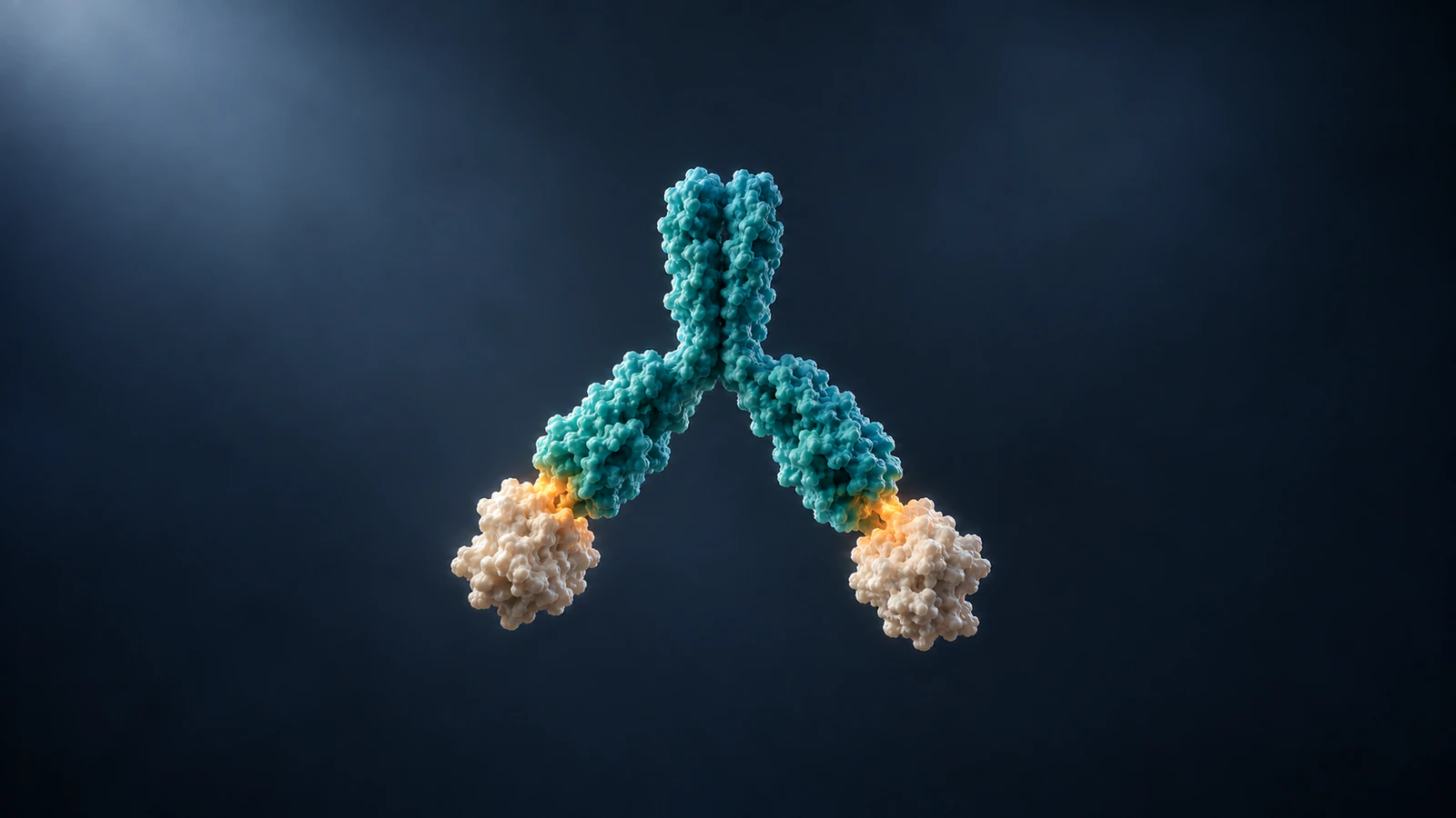
One interaction or combined attachment
Affinity describes the strength of an individual antibody binding site interacting with its target epitope, the specific region it recognizes. Avidity describes the combined attachment in a multivalent interaction, where more than one binding site can contribute. To animate the distinction, keep the individual contact recognizable while showing whether the same antibody has one contact or two.
The useful visual question is therefore not whether an antibody looks more tightly glued to a surface. It is which contacts exist, which remain possible and what evidence supports the comparison. A brighter glow or a longer pause can direct attention, but neither measures binding strength.
This guide develops a conceptual IgG example you can plan in Animiotics. It uses an explicit contact record and a six-shot storyboard rather than an invented affinity score. The proposed sequence is an editorial example, not a reported simulation or a claim that an AI-generated scene is scientifically validated.
Keep the antibody parts identifiable
An IgG antibody has two antigen-binding arms, called Fab regions, connected through a flexible hinge to an Fc region. RCSB PDB-101 illustrates this architecture in its antibody overview. The two tips provide the contact locations in our example; the Fc stem gives the viewer a stable orientation cue.
Build the cast before describing movement: one intact antibody and two separate antigen objects. Give the antibody a single persistent identity even if its arms move. Do not let a close-up quietly replace it with two independent fragments, because that would change what the viewer is being asked to understand.
An isolated Fab can be useful for the first shot, provided the caption identifies it as a fragment. When returning to the intact molecule, make the change of representation explicit. A cut with a short explanation is clearer than a transformation that appears to manufacture a second binding arm.
Explain affinity at one contact
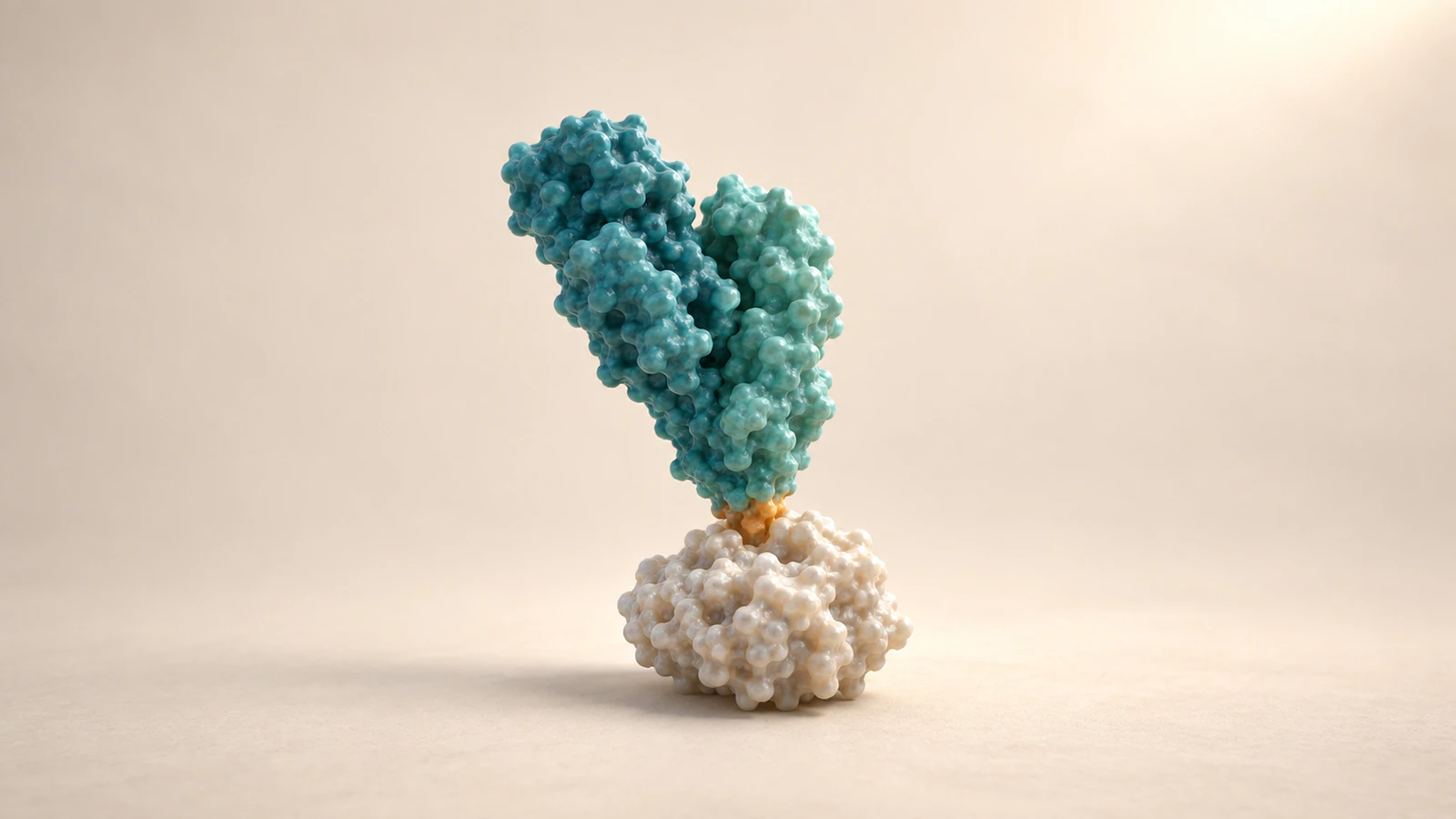
The NCBI MeSH definition of antibody affinity concerns binding strength between an antibody and an antigen determinant. For a teaching shot, isolate the binding tip and the corresponding epitope. Keep enough surrounding surface visible to show which object contributes each side of the interface.
Use a restrained accent at the meeting point and keep the rest of both molecules quiet. The accent means “look here,” not “this bond has a measured energy.” Avoid a permanent weld, a magnet icon or an elastic cord unless you explicitly introduce it as a visual analogy with limits.
If your project includes an experimental affinity value, present it with its source and assay context outside the molecular illustration. Do not assign a number because the objects fit neatly. An attractive interface is a design choice until it is grounded in appropriate structural and experimental evidence.
Show avidity without doubling affinity
For the next shot, widen the camera to include both arms of the same antibody. Show a second accessible epitope and then a second contact. This communicates the change from one attachment to a multivalent configuration without recoloring or enlarging the first contact to imply that its intrinsic affinity has doubled.
Use the phrase “two contacts are now present” in your working notes. It is precise enough to guide the animator and modest enough to avoid a numerical conclusion. Avidity is not an arithmetic instruction to multiply a single-site value by the number of arms.
The screen should let a reader trace an unbroken path from one bound tip through the hinge to the other. If the hinge disappears behind the antigens, choose another view. Otherwise the image may read as two unrelated antibodies, even when your underlying object list is correct.
Make the geometry a condition
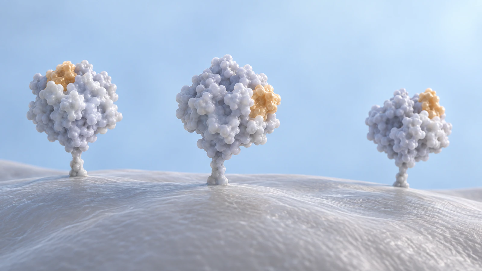
Two available arms do not guarantee two simultaneous contacts. Antigen placement and access matter. A 2021 IgG1 nanocaliper study tested spacing using engineered coiled-coil antigens; its results belong to that experimental system, not to a universal spacing rule for every antibody.
For a conceptual comparison, hold the antibody design constant and change only the position or accessibility of the second target. In one setup, the free tip can reach it without passing through another object. In the other, the second site is visibly unavailable. Describe these as illustrative arrangements rather than experimentally measured separations.
Do not stretch the Fab arm until it reaches. If the scene only works by distorting the protein, the geometry is telling you that the storyboard needs a different arrangement. Preserve the mismatch as a teaching case instead of hiding it with a camera move.
Write a contact record
Before animating, give every shot four short entries: the objects present, the number of antibody–antigen contacts, the available unoccupied site and the evidence level. This record separates a biological claim from a decision about framing. It also gives collaborators a compact way to report a defect.
For this example, the evidence level is “conceptual.” No named antibody, antigen sequence or measured trajectory is assigned to the geometry. If you later substitute a specific molecular system, revisit all four entries rather than keeping the same motion and changing only the caption.
Use the record during review: “Shot four has one remaining contact, but the caption says released” is actionable feedback. “Make the binding clearer” leaves the creator guessing about whether the problem is the camera, the contact point or the underlying claim.
| Shot state | Contact count | What the viewer should see |
|---|---|---|
| Unbound | Zero | Both binding tips clear of their targets |
| First attachment | One | One occupied tip and one visibly free tip |
| Bivalent arrangement | Two | Both contacts on the same intact antibody |
| Half-bound example | One | One contact remains while the other tip is free |
| Complete release | Zero | Neither tip remains attached |
Plan six readable shots
The storyboard below uses presentation beats, not biological time. Decide the length of each beat after checking whether a first-time viewer can identify the relevant contact. A held frame is an opportunity to inspect the scene, not evidence that a complex survives for that duration.
Keep a consistent camera direction through the contact changes. Introduce any close-up before the important event begins and return to a recognizable wide view afterward. For continuity choices, use the same principles described in Molecular Color Continuity.
- Establish the cast: show one intact antibody and two antigens with all parts separated.
- Inspect one tip: use a clearly identified close-up to locate the epitope and binding region.
- Create the first contact: keep the other Fab arm in view or clearly account for it.
- Reveal the second contact: widen enough to trace both arms back to the same hinge.
- Show a half-bound example: release one tip while leaving the other visibly attached. This is a chosen explanatory sequence, not a measured pathway.
- End with the geometry contrast: return to the starting arrangement and show a second target that cannot be reached without distortion.
Keep evidence separate from illustration
A 2026 cryo-electron tomography study examined the P17 and S309 antibodies with SARS-CoV-2 spikes and observed inter-spike binding and different higher-order arrangements. Cryo-electron tomography reconstructs three-dimensional information from electron-microscopy views. The study provides a specific structural context, not permission to label any Y-shaped bridge as a reconstruction of those antibodies.
Our storyboard deliberately avoids reproducing that experiment. Its antigens are generic, its positions are selected for readability and its motion is authored. If you want to explain the published system, use the paper’s actual conditions and structural evidence and have the resulting interpretation reviewed.
For a conceptual article, say exactly what the pictures are: independently generated illustrations of contact configurations. They are not sequential snapshots of one measured molecule. The distinction between authored transitions and scientific trajectories is developed further in Protein Morph vs Simulation.
Build the comparison in Animiotics
Animiotics lets you start with a written scene description and request changes afterward. Begin with the cast and the permitted contacts rather than a dramatic camera direction: “Create a conceptual intact IgG with two identifiable Fab arms and one Fc stem. Show two separate antigens. Keep each object distinct and make the two binding tips easy to inspect.”
Next describe the intended comparison: “Show one tip contacting an antigen while the second tip remains free. Then show a separate, compatible two-contact configuration. Keep the antibody’s identity and proportions consistent. Do not imply a measured binding strength or a universal distance.” This is a proposed starting prompt, not a tested guarantee of the output.
Inspect the resulting scene from multiple angles before directing motion. The product documentation describes timeline review and editing controls; use them to check the beginning, contact changes and final state. Account and workspace options determine available outputs. Scientific interpretation still needs your review regardless of how the scene was generated.
Review the half-bound frame
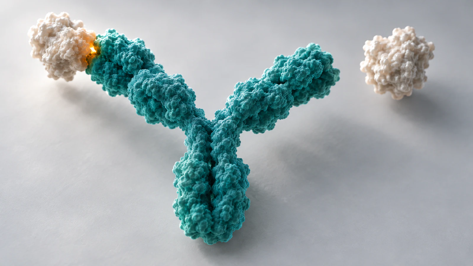
The most useful pause point is often the moment after one contact disappears. Can the viewer identify the remaining attachment without reading the caption? Does the free arm have a visible gap, or is it merely hidden behind an antigen? Is the whole antibody still connected?
Use a second camera angle as a diagnostic view, even if it never appears in the final movie. A silhouette can hide an intersection that becomes obvious from above. Check the actual export as well as the editor view, because a tighter crop can conceal the free tip or the remaining contact.
Finally read the narration against the contact record. Replace “the antibody releases” with “one arm disengages” when one attachment remains. Reserve complete release for a zero-contact frame. That small wording change prevents a polished visual from teaching the wrong event.
FAQ
What is the difference between affinity and avidity?
Affinity concerns one binding-site interaction with an epitope. Avidity concerns the combined attachment in a multivalent setting; an animation should show the contact configuration without pretending to calculate its strength.
Does an IgG always bind with both arms?
No. The second site must be accessible in a compatible arrangement. Show a one-contact state when that is the intended example rather than forcing both arms into contact.
Can two contacts be shown as twice the affinity?
No. Counting contacts is a useful visual check, but it does not provide a numerical affinity or avidity value. Keep measurements tied to the relevant experiment.
Can a generated antibody surface represent a specific structure?
Only if that representation is appropriately grounded and verified. The conceptual illustrations here are not coordinate-derived models of a named antibody or antigen.
Can I use this storyboard in Animiotics?
Yes, use it as a scene-planning and review framework. Describe the objects and contact states, inspect the generated scene and request corrections; the framework does not guarantee scientific accuracy or a particular generated result.
Create a two-contact explanation
Try this focused exercise in Animiotics: create an intact antibody with one occupied tip, then build a clearly distinguished two-contact view. Keep the contact record beside your draft and ask a colleague to identify each state without narration. Refine the framing until the difference is visible before adding a longer mechanism story.