Why Spatial Biology Animation Services Matter
Spatial biology animation services turn tissue maps, biomarker patterns and cell neighborhood evidence into visual stories that buyers can understand without studying every assay output. Spatial biology is powerful because it keeps cells in context. It shows where a signal appears, which cell types sit near each other, how a disease niche is organized and why a platform can see biology that bulk assays miss.
That context is also why the communication challenge is hard. A spatial dataset can contain tissue morphology, segmentation, marker intensity, gene expression, cell typing, neighborhood statistics and clinical interpretation. When all of that appears at once, the story becomes a dense technical artifact rather than a commercial explanation.
Animiotics builds spatial biology visuals for biotech, pharma, diagnostics and research teams that need platform science to work in investor decks, websites, conference talks and partner conversations. The goal is not to flatten the science. The goal is to make the tissue context visible, make the buyer question obvious and connect spatial evidence to platform value. That clarity is often what moves a technical audience toward action.
- Show tissue architecture as the central claim instead of background decoration.
- Translate biomarker patterns into clear mechanism and platform evidence.
- Help buyers understand why spatial context changes the biological answer.
Start With The Spatial Biology Buyer Question
The strongest spatial biology visual starts with the decision the audience needs to make. A pharma partner may ask whether a platform can identify response niches. A diagnostics buyer may ask whether a biomarker pattern supports patient selection. A research audience may ask how spatial transcriptomics, multiplex imaging or proteomics data supports a mechanism. Each question needs a different visual hierarchy.
A useful brief separates assay detail from communication intent. The assay detail might include marker panels, coordinates, cell labels, tissue regions, gene modules or morphology. The communication intent is the claim the viewer should retain after ten seconds. For example: this immune exclusion pattern explains resistance, this biomarker gradient identifies a responsive niche or this platform links cell state to tissue structure.
This article complements Animiotics posts on https://animiotics.com/blog/spatial-proteomics-visualization-services-how-to-turn-marker-panels-tissue-context-and-platform-evidence-into-clear-buyer-ready-visuals/ and https://animiotics.com/blog/how-to-choose-spatial-transcriptomics-visualization-software-for-clear-tissue-atlas-stories/. Spatial biology animation services go further by turning static assay outputs into a guided visual sequence.
- Define the audience decision before choosing the render style.
- Pick one spatial claim for the hero asset.
- Use assay detail only where it supports trust and comprehension.
What To Visualize In A Spatial Biology Story
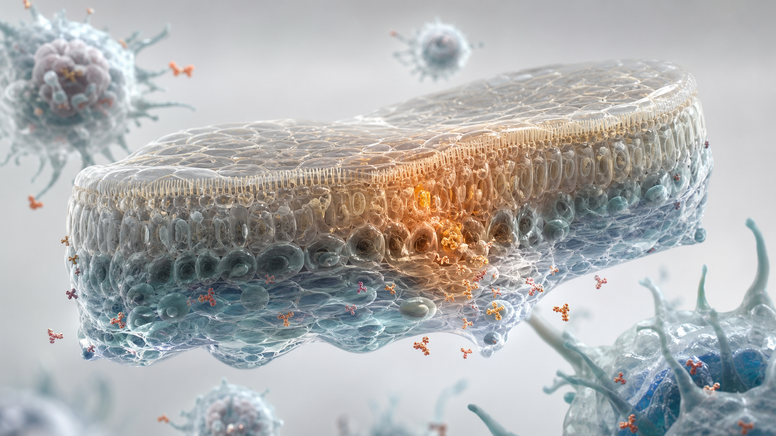
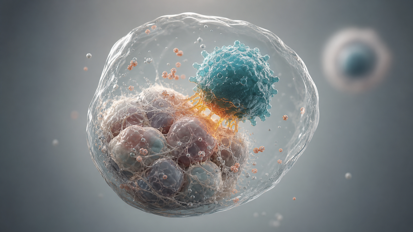
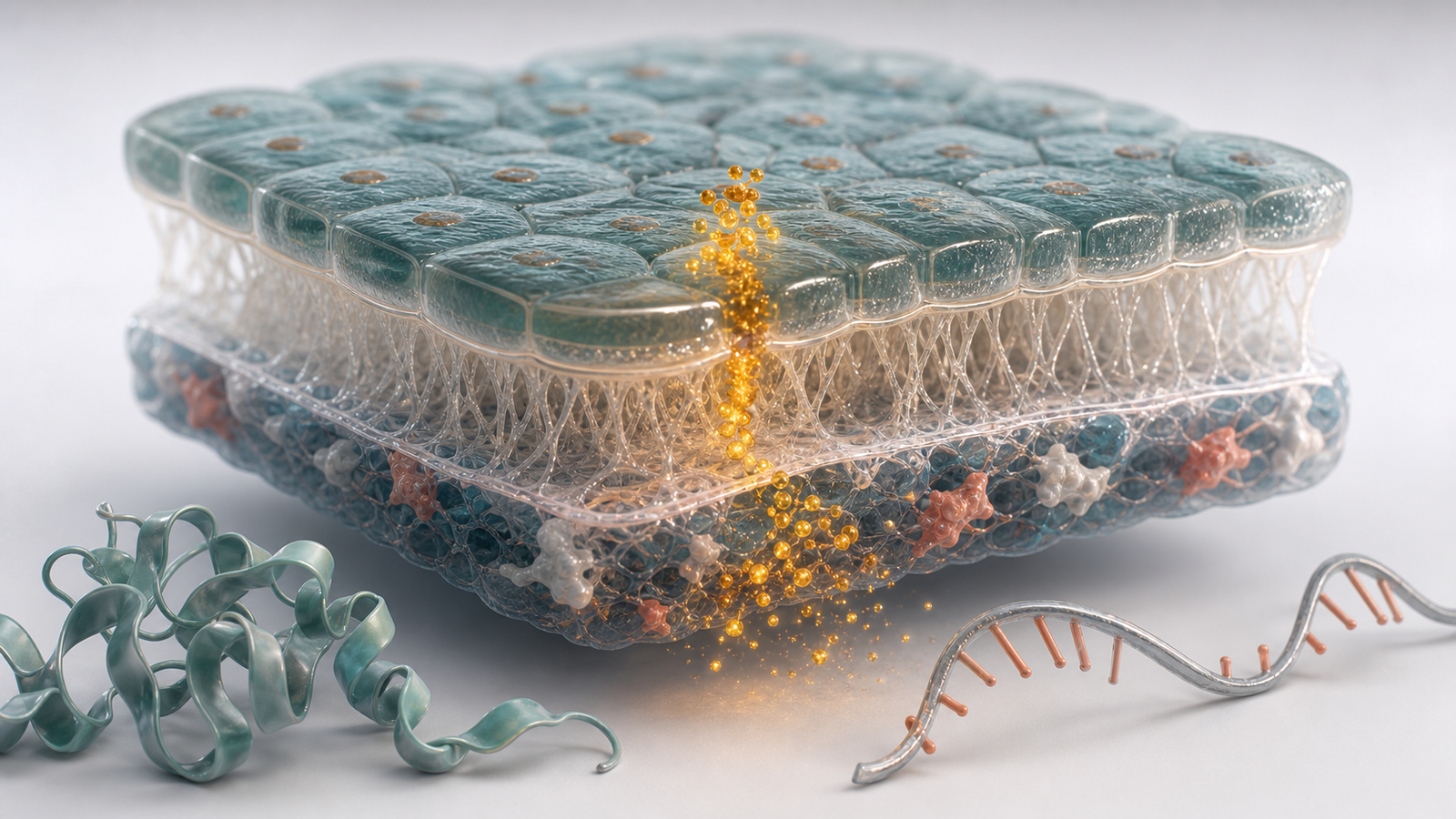
Spatial biology stories usually work best when they move from tissue context to cell neighborhood to molecular signal. The opening view can establish the tissue region, disease niche or sample type. The second view can reveal the cell population or boundary that matters. The third view can show a biomarker pattern, gene module, receptor signal or immune interaction that explains the platform insight.
The visual language should reduce cognitive load. Tissue volumes help non-specialists see context. Transparent cell forms can reveal neighborhoods without hiding structure. Subtle marker particles or signal gradients can show evidence without becoming an infographic. A few supporting molecular forms can connect tissue-scale biology to protein, RNA or membrane mechanisms.
Teams working on oncology, immunology, neurology, fibrosis and inflammatory disease often need this bridge. Related guidance on https://animiotics.com/blog/tumor-microenvironment-visualization-how-to-explain-immune-contexture-drug-delivery-and-oncology-platform-evidence-clearly/ and https://animiotics.com/blog/single-cell-multiomics-visualization-how-to-turn-rna-atac-and-protein-data-into-clear-platform-stories/ covers adjacent mechanism and multiomics stories. Spatial biology animation can unify those signals inside a tissue-aware narrative.
- Tissue morphology for biological context.
- Cell neighborhoods for proximity and interaction.
- Biomarker gradients for mechanism, response and platform differentiation.
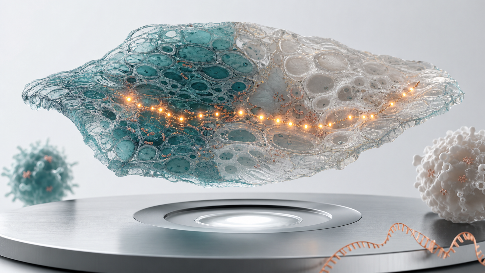
Useful Deliverables For Spatial Biology Teams
Different commercial moments call for different outputs. A website hero needs one elegant tissue signal that says the platform sees biology in context. An investor deck may need a short sequence from sample to spatial map to actionable insight. A partner presentation may need a defensible visual that shows how the assay identifies a niche, stratifies patients or supports target biology.
A practical spatial biology package often includes a cover render, three to five section figures, a short animation loop and a reusable tissue scene system. Reusability matters because spatial programs evolve as marker panels, cohorts and disease questions change. If camera angles, cell materials, tissue regions and biomarker colors are consistent, the team can update the science without rebuilding the visual identity every time.
The table below maps common spatial biology communication needs to assets that usually work well.
| Need | Best visual asset | Why it works |
|---|---|---|
| Explain assay architecture | Tissue section render with signal path | Shows how measurement stays connected to context |
| Show cell niche biology | Neighborhood scene with highlighted contact zone | Makes proximity and interaction easy to see |
| Support biomarker value | Marker gradient or response niche sequence | Connects data pattern to a clinical or platform claim |
| Pitch a spatial platform | Reusable tissue visual system | Turns multiple datasets into one coherent story |
How To Keep Spatial Biology Visuals Scientifically Honest
The main risk in spatial biology visualization is over-simplifying the data until the biology looks cleaner than it is. A polished tissue render can imply a sharp boundary, direct causality or uniform marker pattern when the source data is more probabilistic. A careful workflow keeps assay method, data type, confidence and simplification notes visible during review.
Scientific review should happen before final polish. The review package can include source images, segmentation notes, marker definitions, cell type labels, region annotations and a short description of what each scene is allowed to claim. This helps biologists, computational scientists and commercial leads agree on the story before materials, lighting and camera work make the scene feel final.
The final asset can still be elegant. The point is to simplify responsibly. Show the tissue region that matters, emphasize the cell niche that supports the claim and leave out data layers that do not help the viewer understand the decision. Spatial biology animation services are most valuable when they make complexity legible without pretending uncertainty does not exist.
- Document which dataset supports each visual claim.
- Separate measured signal from interpreted mechanism during review.
- Use stylization to guide attention, not to exaggerate certainty.
Where Spatial Animation Adds Platform Value
Static spatial figures can show a result, but animation can show why the result matters. Motion can guide the viewer from tissue overview to cell neighborhood to molecular signal. It can reveal a marker gradient, immune boundary, stromal niche or vascular pattern in a sequence that mirrors how the scientific team reasons through the evidence.
This matters commercially because spatial biology platforms are often sold on insight, not only on measurement. Buyers want to know what the platform sees that other methods miss. A clear animation can connect sample preparation, spatial readout, computational interpretation and biological decision in one coherent story. That is valuable for business development, fundraising, launch campaigns and conference presentations.
A reusable animation system also supports repeated communication. The same tissue materials, camera grammar, biomarker palette and cell forms can be used for multiple disease programs. That consistency helps a platform feel credible and helps audiences remember the scientific position.
- Use motion to reveal spatial hierarchy instead of adding visual noise.
- Connect tissue context to biomarkers, cell niches and actionability.
- Build a visual system that supports multiple programs and datasets.
FAQ About Spatial Biology Animation Services
What inputs are useful for spatial biology animation services?
AHelpful inputs include microscopy images, segmentation outputs, marker panels, tissue region notes, cell type definitions, gene or protein signatures, existing figures and a short explanation of the claim. Screenshots are useful for intent, but source files and review notes make the final asset more accurate.
Can spatial visuals be used before the full dataset is public?
AYes, if the claims are handled carefully. A stylized visual can communicate platform capability, assay logic or disease context without revealing proprietary data. The review process should define what is measured, what is representative and what must remain generalized.
How many biomarkers should appear in one visual?
AFor a buyer-facing hero render, one or two signals are usually strongest. For a technical sequence, three or four layers can work if the visual hierarchy keeps the main signal readable.
Do these visuals replace scientific figures?
ANo. They translate spatial biology into communication assets. A technical figure may still need exact markers, scales, coordinates and methods. A commercial render or animation helps the audience understand why the spatial result matters before they study the full evidence.
Ready To Turn Spatial Biology Into Buyer-Ready Visuals
If your team has spatial omics, multiplex imaging, tumor microenvironment or tissue atlas evidence that is hard to explain outside the core science group, Animiotics can help turn it into clear renders, figures and animation assets. We can build a visual system around tissue context, biomarkers, cell niches, assay workflow or platform differentiation so the story works for scientific review and commercial communication.
Start with the buyer question, then bring the dataset context, current figures, marker definitions and program goals. Animiotics can help shape the story, design the tissue visual system and produce assets for a website, deck, conference talk or partner discussion. To start a project, visit Animiotics or use /pricing?from=blog.
- Best fit: biotech, pharma, diagnostics, platform and research teams with spatial evidence to explain.
- Typical outputs: cover render, section figures, short animation loops and reusable tissue scenes.
- Primary goal: make spatial biology commercially useful without weakening the science.