Why Organ On Chip Platforms Need A Visual Story
Organ on chip animation services matter because microphysiological systems can be hard to understand from static schematics alone. A platform may combine engineered tissue, microfluidic perfusion, barrier biology, sensor readouts and drug exposure in one compact device. Scientists may understand the model, but investors, partners and cross-functional buyers often need a clearer path from chip architecture to translational value.
The central challenge is not just showing a tiny device. It is explaining why the model behaves more like relevant biology than a flat culture plate and why that difference supports a better decision. A strong animation can show cells organizing in a channel, media flow creating shear, a compound entering the system, tissue response changing over time and assay evidence connecting back to a specific development question.
For commercial communication, that story has immediate value. Buyers want to know what the chip measures, where the biology is credible, what decision the assay supports and how the platform could reduce uncertainty before clinical work. A polished visual gives those questions a shared frame before the team moves into detailed validation data.
- Use organ on chip visuals when device design, tissue biology and assay evidence are scattered across too many slides.
- Show the model as a living experimental system rather than a decorative microfluidic diagram.
- Connect the physical chip to a concrete buyer question such as toxicity, efficacy, barrier transport or disease response.
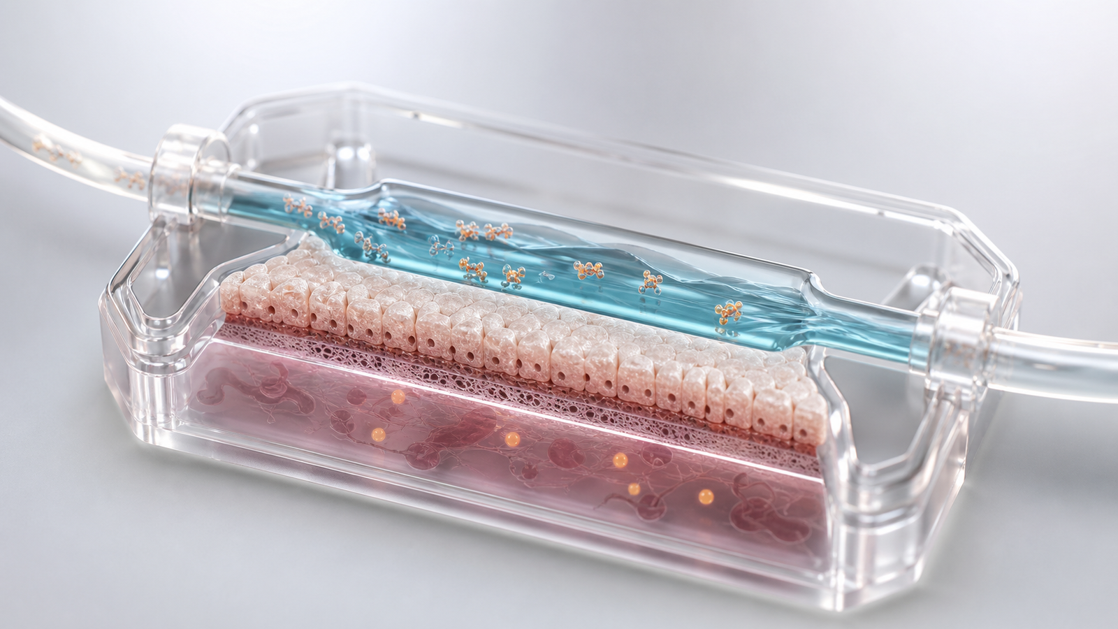
Start With The Microphysiology
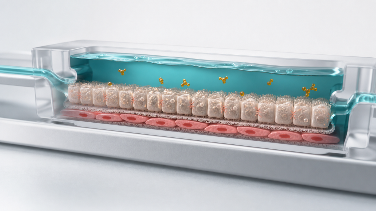
The first scene should make the biological model obvious. A gut barrier chip, liver toxicity chip, lung airway chip, tumor microenvironment chip or vascular model each carries a different communication burden. The viewer should understand which tissue is represented, how the cells are arranged and why the flow or mechanical context changes the interpretation of the assay.
A useful opening usually centers on one hero chip region instead of a full device exploded view. The frame can reveal a transparent microchannel, a structured epithelial or endothelial layer, a matrix zone and a controlled flow path. Only a few supporting molecules, immune cells or stromal cues should be added. The goal is to create a readable mental model that can survive later discussion of endpoints and validation.
This is where premium 3D rendering outperforms generic line art. Subtle membrane textures, physically plausible fluid materials and clear depth cues help the scene feel engineered without becoming cluttered. The visual should look like a credible scientific model, not a toy device or a fake dashboard.
- Identify the tissue type, channel geometry and assay context before adding data cues.
- Use one clear chip region as the hero subject so the viewer understands the platform quickly.
- Keep the scene grounded in plausible cell and material behavior.
| Platform feature | What the visual should clarify |
|---|---|
| Perfused channel | How flow exposes tissue to media, drug or immune components |
| Barrier layer | How cells form a functional interface that can be challenged or measured |
| Disease model | How the chip reproduces the relevant biology without overclaiming clinical equivalence |
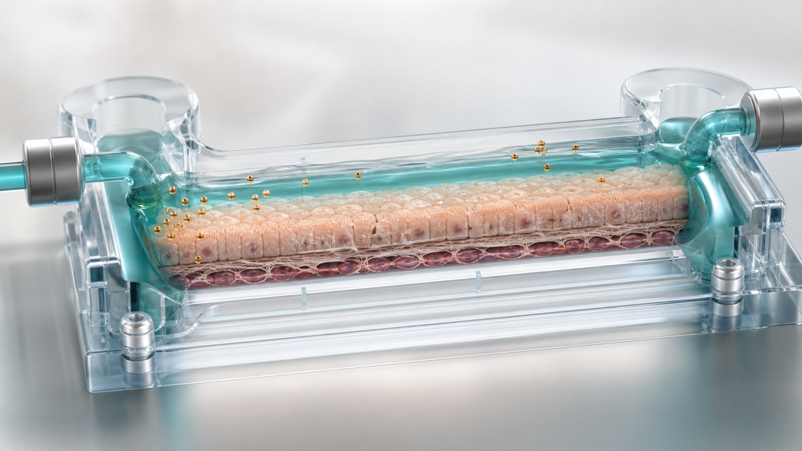
Turning Assay Design Into Buyer-Ready Animation
Buyer-ready organ on chip animation does not need to show every protocol step. It needs to explain the decision the assay supports. A drug discovery team may need to show whether a compound crosses a barrier. A safety team may need to show tissue injury or functional decline. A platform company may need to show how the same device architecture can support multiple disease models while preserving a consistent experimental logic.
The strongest visual sequence starts with setup, then shows perturbation and response. First, the chip establishes tissue organization and flow. Next, the compound or biological trigger enters the system. Then, the tissue changes in a way that maps to the endpoint: permeability, contractility, cytokine release, morphology, viability or downstream biomarker response. Finally, the animation connects the result to the buyer's development question.
This structure pairs well with existing Animiotics guidance on preclinical pharmacology animation services because both stories translate model evidence into decision confidence. It also connects with translational biomarker visualization services when the chip output supports patient selection, response prediction or mechanism validation.
- Frame each visual beat around the decision the assay informs.
- Separate setup, perturbation and biological response so the story stays easy to follow.
- Use the same material language across still figures, website renders and full animation.
Drug Response, Toxicity And Translational Confidence
Drug response is where organ on chip visuals become commercially persuasive. A program may have promising cell data, but buyers still need to understand whether the model adds relevant context. Does flow change exposure? Does tissue architecture reveal a response that a simpler assay misses? Does the chip help interpret toxicity, efficacy or patient-specific behavior before a costly development step?
A clear animation can show these relationships without pretending the model replaces clinical evidence. Low exposure may leave the tissue largely unchanged. A pharmacologically relevant exposure may shift a measurable response in the intended direction. Excess exposure may produce barrier disruption, altered morphology or injury cues. That visual logic helps mixed audiences see how the assay supports dose selection, candidate ranking or safety assessment.
Organ on chip communication should also respect uncertainty. If a model is validated for a specific endpoint, the visual can highlight that endpoint. If the platform is exploratory, the animation should emphasize hypothesis testing and evidence generation rather than direct prediction. Credibility grows when the visual makes the platform's strengths clear while avoiding claims the data cannot carry.
- Show drug response as a relationship between exposure, tissue state and measured endpoint.
- Avoid implying full clinical prediction when the model supports a narrower decision.
- Use restrained biological cues to show toxicity or efficacy without turning the frame into an infographic.
| Use case | Visual emphasis | Commercial takeaway |
|---|---|---|
| Barrier transport | Compound movement across a structured cell layer | The model can clarify access and permeability |
| Toxicity assessment | Tissue stress, morphology change or functional decline | The assay can flag risk before later development work |
| Disease response | A controlled perturbation with a measurable tissue reaction | The platform can support candidate selection and mechanistic confidence |
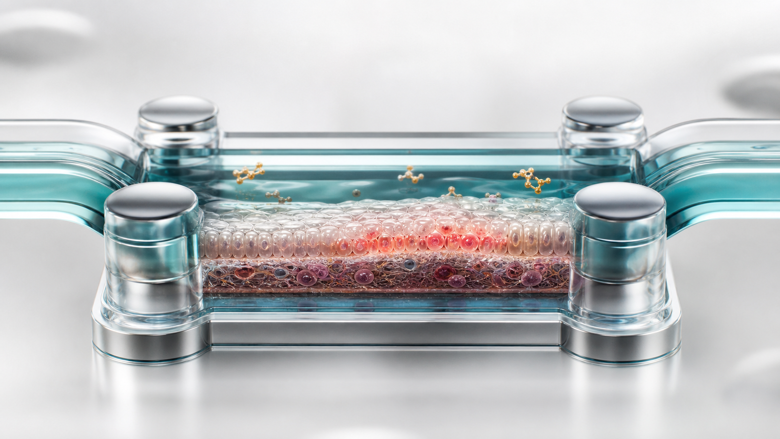
Visual Choices That Build Platform Trust
Organ chip visuals can lose trust quickly when they become too glossy or too abstract. A busy collage of channels, floating cells, glowing beads and fake interface panels may look high tech, but it can make the platform feel less specific. Trust comes from a clean scientific scene where the viewer can identify the tissue, the channel, the flow path and the biological event.
Materials should feel lab-grade and physically plausible. Microfluidic structures can be translucent without looking like candy. Fluid should have a subtle surface and believable volume. Cells should be simplified enough for readability while retaining membrane, matrix or tissue character. Molecular cues should support the assay story instead of filling empty space.
Color also matters. A restrained biotech palette of muted teal blue, pearl white, silver gray, warm amber and sparse coral accents keeps the image premium and consistent with modern scientific rendering. The scene should avoid labels, fake UI, dashboards, chart overlays, logos and visible text. The visual should feel like a handcrafted Blender or Maya science render built to explain the platform.
- Use one clear biomolecular or tissue-chip hero subject with only a few supporting forms.
- Favor plausible channels, membranes, tissue volumes and assay cues over decorative particles.
- Keep text, labels, fake interfaces and chart overlays out of the image.
Where Organ On Chip Visuals Create Leverage
Organ on chip animation services can support many buyer-facing moments. On a website, a hero render can explain the platform in seconds. In a pitch deck, a short sequence can show how the assay connects to development risk. In a partnering meeting, the same visual language can explain model setup, endpoint logic and platform expansion without rebuilding the story from scratch.
The assets are especially useful when different stakeholders care about different layers of the platform. A scientist may ask about cell sourcing, validation and endpoint sensitivity. A business development audience may focus on model relevance and partnership fit. An investor may need to understand why the platform can support a pipeline or service business. A coherent visual system lets each audience enter the same story at the right level.
For teams building a broader content strategy, organ chip visuals can sit beside mechanism of action animation, biomarker figures, preclinical model renders and platform storyboards. The result is a reusable communication system that helps scientific, executive and commercial teams explain one model consistently across channels.
- Use still renders for web pages, conference materials and investor slides.
- Use short animation sequences for assay workflow, drug response and platform value.
- Build reusable scenes so future indications and endpoints can share the same visual language.
FAQ
What are organ on chip animation services?
AOrgan on chip animation services create still images, 3D renders and motion sequences that explain microphysiological systems, tissue-chip assay design, drug response and platform value for biotech or pharma audiences.
Who needs organ on chip visuals?
APlatform companies, drug discovery teams, preclinical groups, translational science teams, CROs and pharma innovation teams use organ chip visuals when they need to explain a complex model to buyers, investors, partners or internal decision makers.
Can an organ chip animation stay scientifically accurate?
AYes. The animation should be scoped around the model's validated biology, assay endpoints and intended decision. A credible visual can show flow, tissue architecture and response while avoiding claims that go beyond the evidence.
What can Animiotics build from an organ chip story?
AAnimiotics can build website hero renders, pitch deck figures, section images, conference visuals, assay workflow animations and reusable 3D scenes that support organ on chip platform communication.
Ready To Build An Organ On Chip Visual
A strong organ on chip story starts with the tissue model, the assay endpoint and the decision your audience needs to make. Animiotics can turn that material into premium 3D scientific renders and animation-ready scenes that explain microphysiology, drug response and platform value with the clarity buyers expect.
Start with the chip architecture, tissue context, flow logic, endpoint and commercial moment the asset needs to support. From there, the visual can be scoped as a single hero image, a set of section renders or a full animation sequence. For broader planning, review Animiotics guidance on scientific animation services for biotech and then visit Animiotics to discuss an organ on chip visual for your platform.
- Use organ chip visuals when model relevance is central to buyer confidence.
- Connect device design, tissue biology and assay response in one coherent visual system.
- Plan still renders and animation scenes together so the platform story stays consistent.