Choose the complex before the camera
Biological assembly animation starts with a selection decision: which protein chains belong in the complex you intend to show? Check the deposited assembly, its supporting evidence and the number of displayed subunits before planning motion. A convincing surface render can otherwise turn an incomplete structure or a crystal neighbor into the wrong biological object.
For scientists preparing an Animiotics animation, this is a useful first review. The goal is a scene whose identity survives every camera move, visibility change and close-up. This guide gives you an assembly record, a short inspection storyboard and a concrete archive example to help make that decision.
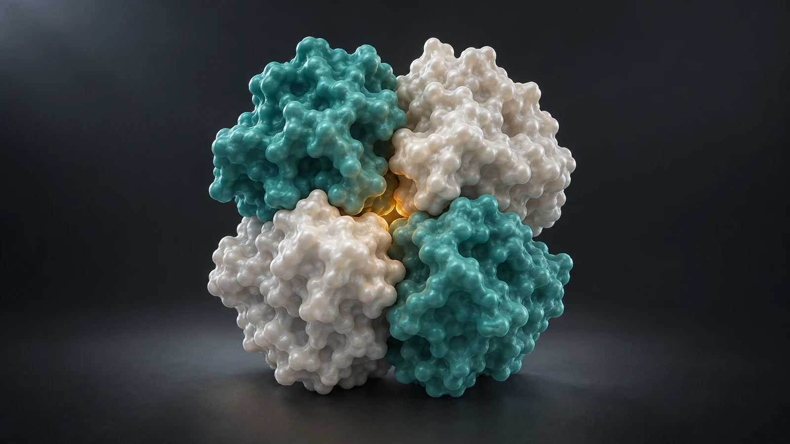
The accompanying images are conceptual CGI illustrations. Their shapes are not generated from PDB coordinates, and they do not depict the named hemoglobin example. Use them to understand the visual decisions; use the archived structure and its documentation to establish an actual molecular arrangement.
Asymmetric unit or biological assembly
In crystallography, an asymmetric unit is the unique portion from which crystal symmetry generates the repeating structure. A biological assembly describes a molecular grouping shown or believed to be functionally relevant. The two can coincide, but an asymmetric unit can also contain only part of an assembly or several copies of it. RCSB explains these possibilities in its biological-assemblies guide.
That distinction changes the first production question. Instead of asking whether the downloaded object looks complete, ask what coordinate selection the download represents. Save both the entry identifier and the selected assembly identifier. A filename alone is a poor substitute for that record.
Keep the original download untouched and make a separate presentation copy. When a reviewer challenges the number of pieces on screen, you can then trace the answer to the source rather than to an undocumented edit. Do this before assigning materials, because attractive colors can make an arbitrary grouping feel intentional.
Count identities and copies separately

RCSB distinguishes an entity, a chemically unique molecule, from an instance, a particular occurrence of that molecule. An assembly groups relevant instances. Two different protein types can therefore appear as four protein-chain copies. Chain identifiers distinguish instances within an entry, but their lettering is not a universal naming system across entries. See the PDB structure hierarchy.
Translate that hierarchy into a small production inventory. Record the molecular type separately from the name you give its displayed copy. If two identical subunits have different screen names, that should help you track them, not suggest different chemistry. Conversely, assigning the same material to every chain must not erase a distinction that matters to the explanation.
Count protein chains and retained non-protein components separately. Then compare the inventory with what is actually visible in the opening frame. A chain hidden behind another may be present in the scene but unavailable to the audience. Adjust the camera or introduce a brief inspection view rather than silently deleting the hidden copy.
A real example: trout hemoglobin
PDB entry 1OUT describes trout hemoglobin I from Oncorhynchus mykiss, not human hemoglobin. Its author-assigned Biological Assembly 1 is a heterotetramer with A2B2 stoichiometry: two copies of each of two protein types. The entry is an X-ray diffraction structure at 2.30 angstrom resolution; the page reviewed here identifies version 1.5.
The RCSB assembly guide uses this entry to illustrate an asymmetric unit containing two protein chains while the biological assembly contains four. The remaining pair is generated using the documented symmetry relationship. This is a case where counting the initially displayed chains could produce the wrong hero complex.
For a teaching exercise, inspect the asymmetric unit and Biological Assembly 1 separately. Write down the protein-copy count before comparing the views, then confirm that your presentation asset matches the assembly you selected. Keep the organism and entry visible in your working notes so the exercise does not become an unsupported claim about a human protein.
This example establishes a selection problem, not an animation of oxygen binding or a path between functional states. Those would require additional evidence and a different brief. A structure chosen to teach assembly membership should not acquire a mechanistic story simply because its name is familiar.
Keep an assembly record
Use one compact record that travels with the scene. It should be short enough to read during review but specific enough to reproduce the selection. The table below is a proposed production framework, not a new archive standard.
Resolve disagreements in this record before changing the scene. If the scientific reviewer expects four chains and the artist sees two, first compare the selected assembly and visibility settings. Adding two convenient duplicates by eye may fix the count while leaving the spatial arrangement wrong.
A useful handoff includes the source file, the reviewed selection and one neutral overview image. Keep the overview free of dramatic depth of field so that the reviewer can identify the pieces. The polished cover is a communication asset; the neutral overview is a check on what the asset contains.
| Record | What to retain |
|---|---|
| Identity | PDB entry, organism, construct and downloaded version |
| Selection | Assembly ID, assignment information and source file |
| Membership | Protein types, copy count and retained cofactors |
| Display changes | Hidden pieces, color mapping and inspection offsets |
| Evidence boundary | What this selection supports and what remains unresolved |
Use documented placement
The PDBx/mmCIF archive format records assembly-generation instructions. The pdbx_struct_assembly_gen category associates an assembly identifier with component identifiers and an operation expression describing the required transformations. This is why a supported arrangement should come from the assembly data rather than manually placing copies until the silhouette seems plausible.
For a production handoff, retain either the downloaded assembly coordinates or the precise procedure used to generate them. If a preparation tool creates new copy names, record their relationship to the source identifiers. Do not assume that the letters used in a viewer are interchangeable with every identifier field in the file.
After preparation, reopen the exported asset and compare it with the reviewed reference view. Verify membership, relative placement and retained components again. This check catches a practical class of error: the source selection was correct, but an export omitted a hidden object or an artist duplicated something for an earlier layout experiment.
Choose the display representation after that check. Our guide to protein surface versus ribbon helps match the representation to the question without changing which pieces belong in the assembly.
Treat crystal contacts as a question
A nearby protein in a crystal is not automatically another functional subunit. Assembly assignments can involve interpretation. PDBePISA explores molecular interfaces and calculates probable assemblies and related properties. Those calculations are useful evidence to examine, but a visually convincing interface is not by itself a complete biological argument.
When possible assignments disagree, put the alternatives into the scientific review instead of choosing the one with the strongest silhouette. Ask which state the article or animation needs to describe and which evidence supports that state. Save the decision and any remaining limitation with the source record.
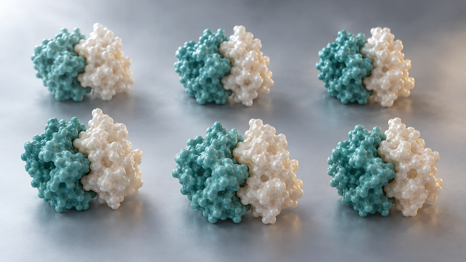
For an illustrative crystal-context shot, show repetition as context and keep the selected complex visually traceable. Avoid turning the entire repeated array into one moving object called the protein. The contextual image here uses spaced conceptual pairs to teach that distinction; it is not a crystallographic packing model or a measured lattice.
Build a five-shot inspection
Try this storyboard before adding a biological event. It answers one narrow question: can a viewer identify the selected assembly and track its members? The times are deliberately unspecified because reading pace depends on the audience, narration and final display size.
Keep colors stable across the sequence. If the camera rotates while a copy becomes visible, give the audience a pause before changing representation too. A simultaneous spin, material change and separation makes it difficult to tell whether the object changed or the viewpoint changed.
In the inspection shot, any separated pieces are displaced for explanation. Call that an exploded view in the caption or narration. It must not resemble evidence that the protein spontaneously dissociates along those paths. Restore the reviewed arrangement before showing an interface in its structural context.
- Establish: hold the complete selected assembly with every major subunit distinguishable.
- Identify: introduce the molecular types while preserving the camera and copy count.
- Inspect: separate displayed copies only if counting or occlusion requires it; disclose the editorial displacement.
- Restore: return the same copies to the reviewed arrangement and hold for comparison.
- Focus: approach one interface while preserving enough surrounding structure to locate it.
Review motion independently
Correct membership does not validate a proposed motion. An assembly selection tells you which arrangement you chose to depict; it does not supply a measured association pathway, dissociation rate or order of arrival. Treat any such claim as a separate scientific question with its own evidence.
Write the intended sentence beside each moving shot. For example, an inspection rotation can support 'these are the four displayed subunits.' It cannot support 'these four pieces assemble in this order.' If the sentence exceeds what the source establishes, change the narration, change the motion or gather the missing evidence.
Also distinguish a missing subunit from missing residues within a chain. The first concerns membership; the second concerns how much of a chain has coordinates. Our guide to missing residues covers that separate review, including when omission or an explicitly schematic connector is appropriate.
Keep both checks visible in your review notes. A complete copy count can coexist with unresolved regions, and filling those regions does not settle an assembly assignment. This prevents one successful check from being treated as approval of the whole scientific scene.
A release check for the scene
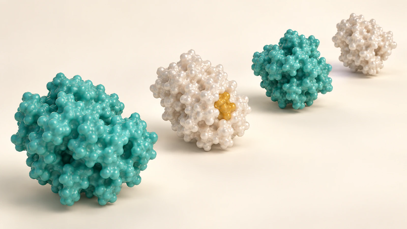
Review the first frame, the most separated inspection frame and the final restored view together. The molecular types and copy identities should remain consistent. If a piece vanishes, determine whether it is occluded, intentionally hidden or absent from the asset; those are different explanations.
Check the smallest intended presentation size as well. Thin seams between subunits may disappear in a thumbnail, especially when adjacent materials have similar brightness. Improve lighting or camera position before exaggerating the physical gap. If you deliberately separate the pieces, retain the exploded-view disclosure.
Finally, read the caption without the animation. It should name the selected subject, explain any display manipulation and state relevant limits. A reviewer who receives only a still should not mistake editorial spacing for an experimental arrangement. Save the approved frame and assembly record with the project so that later revisions have a clear reference.
FAQ
Is the asymmetric unit always the wrong choice?
No. It may match the biological assembly, contain part of it or contain multiple assemblies. Check the specific entry and the purpose of your view before selecting coordinates.
Does a biological assembly always contain several chains?
No. A biologically relevant assembly can be a monomer. Do not add neighbors just to make the scene look more complex.
Can I build the assembly by duplicating a chain manually?
A matching copy count is insufficient. Use the documented assembly coordinates or generation instructions and verify the resulting placement against the selected source.
Does an exploded view show a real dissociation mechanism?
No. Editorially separating pieces helps inspection but does not establish a physical pathway. Disclose the manipulation and restore the supported arrangement for structural claims.
Are the images in this article exact hemoglobin structures?
No. They are conceptual protein-surface illustrations used to explain selection and review. The 1OUT example is documented in the text and must be inspected through its archive record.
Try an assembly inspection in Animiotics
Use Animiotics to create a focused protein-complex animation: establish the whole subject, make its subunits readable and finish on one interface. Prepare the assembly record first, then use it to review what the finished scene communicates. Keep any conceptual geometry or inspection offsets explicit rather than presenting them as experimental coordinates.
Start with that single inspection sequence and assess whether another scientist can follow the membership without your explanation. Once the identity and arrangement are clear, you have a better basis for deciding whether a separate mechanism sequence is justified.