Why siRNA Therapeutic Mechanism of Action Animation Matters
A siRNA therapeutic mechanism of action animation turns a complex intracellular process into a story that investors, partners, clinicians and scientific reviewers can follow in seconds. The biology is specific: a short interfering RNA is delivered to the right tissue, escapes the delivery compartment, loads into the RNA-induced silencing complex and guides sequence-specific cleavage of target mRNA. The commercial challenge is that each step can look abstract if it is shown only as dense pathway art or a static sequence diagram.
For biotech teams, siRNA visuals often need to explain more than RNA interference itself. They must show delivery strategy, tissue tropism, target biology, duration of knockdown, biomarker logic and platform repeatability. A strong animation gives each of those claims a visual place without making the viewer memorize every molecular detail.
The best result is not a decorative explainer. It is a reusable scientific asset system that can support investor decks, business development meetings, conference screens, website pages, manuscript figures and internal program reviews. That is why siRNA MOA work benefits from the same planning discipline used in mRNA vaccine mechanism of action animation and other therapeutic platform stories.
- Use animation to connect delivery, intracellular activity and measurable knockdown.
- Keep the visual hierarchy centered on the therapeutic claim.
- Build reusable scenes that can serve the platform as new targets are added.
Start With Delivery and Cellular Entry
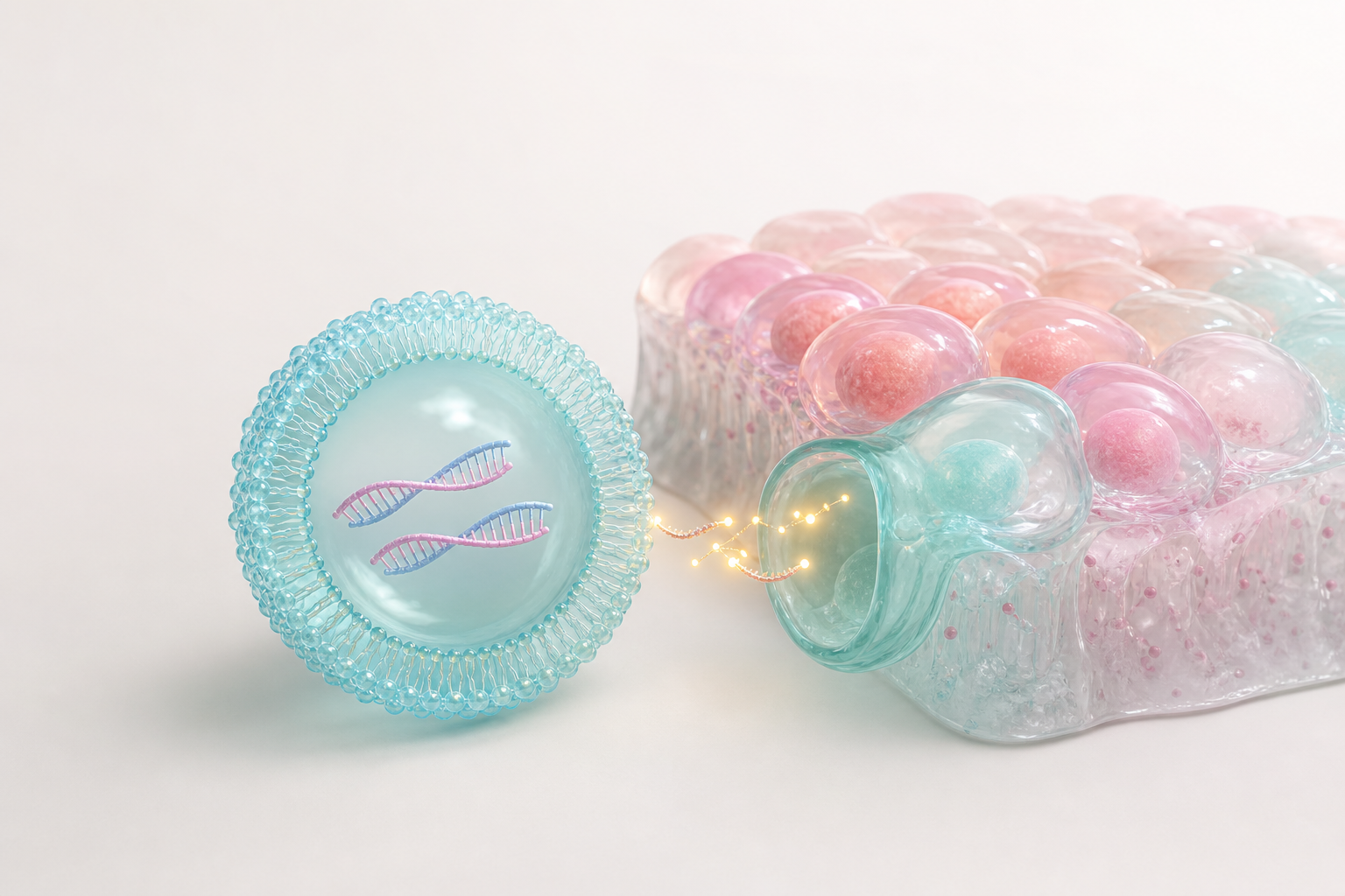
Most siRNA stories begin outside the target cell. The viewer needs to understand how the therapeutic gets close enough to matter before the animation moves into intracellular RNA interference. That may involve a lipid nanoparticle, conjugated ligand, receptor-mediated uptake, local administration or another delivery approach. The visual should make the delivery modality clear without turning the first scene into a crowded pharmacokinetic map.
A practical approach is to show a simplified tissue environment, then isolate the delivery vehicle as it approaches a target cell population. Color can separate carrier, cargo and tissue context. Motion can show binding, uptake and compartment formation. The camera should stay patient enough for a non-specialist audience to understand what is entering the cell and why the cell type matters.
This section should also establish what the animation will not show. If the mechanism depends on a specific ligand, receptor or tissue compartment, that element should be rendered with scientific intent. If the audience does not need subcellular precision at this moment, the first delivery shot can remain clean and conceptual. The goal is orientation before detail.
- Show the delivery vehicle as a distinct object with consistent color and material.
- Use target tissue context sparingly so the carrier remains the hero.
- Reserve high-detail molecular views for the steps that drive the claim.
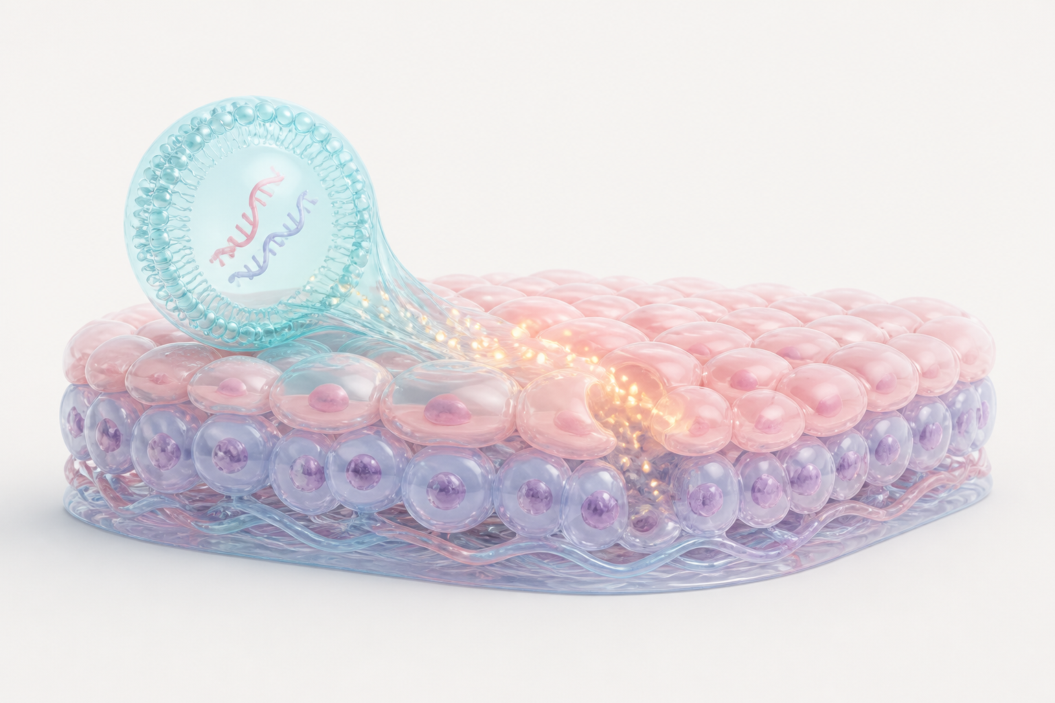
Visualize Endosomal Release Without Overbuilding the Cell
Endosomal release is one of the hardest siRNA steps to explain visually because it sits between cellular uptake and productive cytoplasmic activity. If the animation skips this moment, the viewer may not understand how the cargo reaches RISC. If it overbuilds the endosome, lysosome, cytosol and membrane traffic, the story can become slower than the business use case requires.
A clear siRNA delivery visualization usually benefits from a stylized cross-section. The delivery vehicle can enter a soft translucent compartment, then release paired RNA strands through a controlled opening, color shift or glow. The animation does not need to imply that every particle escapes. It should show the intended productive route and keep the viewer focused on the mechanism the therapy is designed to achieve.
This is also where scientific review matters. Teams should confirm that the release scene does not imply unsupported efficiency, a wrong compartment or a delivery route that conflicts with the platform. A beautiful render that misstates release biology creates more risk than value. A restrained visual that explains the intended step accurately is more useful for diligence and education.
- Use translucency to make intracellular compartments readable.
- Show release as a selective event rather than a universal guarantee.
- Review the delivery route with platform scientists before final animation.
Make RISC Loading the Mechanistic Turning Point
RISC loading is the moment when the siRNA becomes a sequence-guided silencing tool. In a static figure, this step can look like a generic protein-RNA interaction. In animation, it can become the turning point of the mechanism: the duplex arrives, one strand is retained as the guide, the passenger strand leaves and the loaded complex begins scanning for complementary mRNA.
The visual language should be simple. A pearl-like protein complex, a paired blue and pink RNA duplex and a clean guide strand are often enough. The animation can use a slow camera move, a slight opening of the complex and a color transition to indicate activation. The important message is not every atomic contact. The important message is that sequence information is now loaded into a cellular machine.
For platform teams, this step is commercially useful because it connects chemistry and biology. Modifications, potency claims, durability and target selectivity all become easier to discuss when the viewer understands that the guide strand directs the silencing complex. The same scene can later be adapted for different targets, tissues or candidate programs.
- Treat RISC loading as the central mechanism beat.
- Differentiate guide strand and passenger strand with stable colors.
- Avoid showing unsupported structural detail unless the team has evidence.
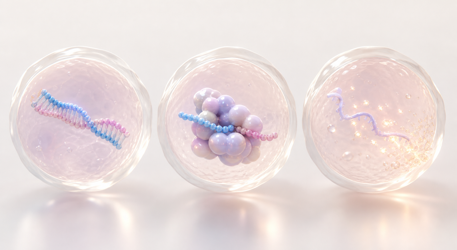
Show Target mRNA Cleavage and Knockdown as Evidence
The payoff of siRNA mechanism animation is target knockdown. After RISC loading, the guide strand directs the complex to complementary mRNA, cleavage occurs and the amount of translated target protein decreases. A clear animation needs to show this change as a biological consequence, not just as a disappearing ribbon.
One useful sequence is to show target mRNA entering the RISC scene, aligning with the guide, separating after cleavage and fading as downstream protein output declines. That can transition into a simplified cellular or tissue readout, such as reduced pathological signal, lower target protein abundance or a cleaner biomarker state. The transition from molecule to evidence helps the audience understand why the mechanism matters.
This logic aligns with broader translational biomarker visualization services. The mechanism is stronger when it connects to assay readouts, pharmacodynamic markers or patient-selection rationale. Animation can make that bridge visible without pretending that one render replaces the data.
- Show target mRNA recognition before cleavage so specificity is clear.
- Connect molecular silencing to a measurable biological readout.
- Keep efficacy language aligned with the actual program evidence.
Build a Reusable siRNA Platform Visual System
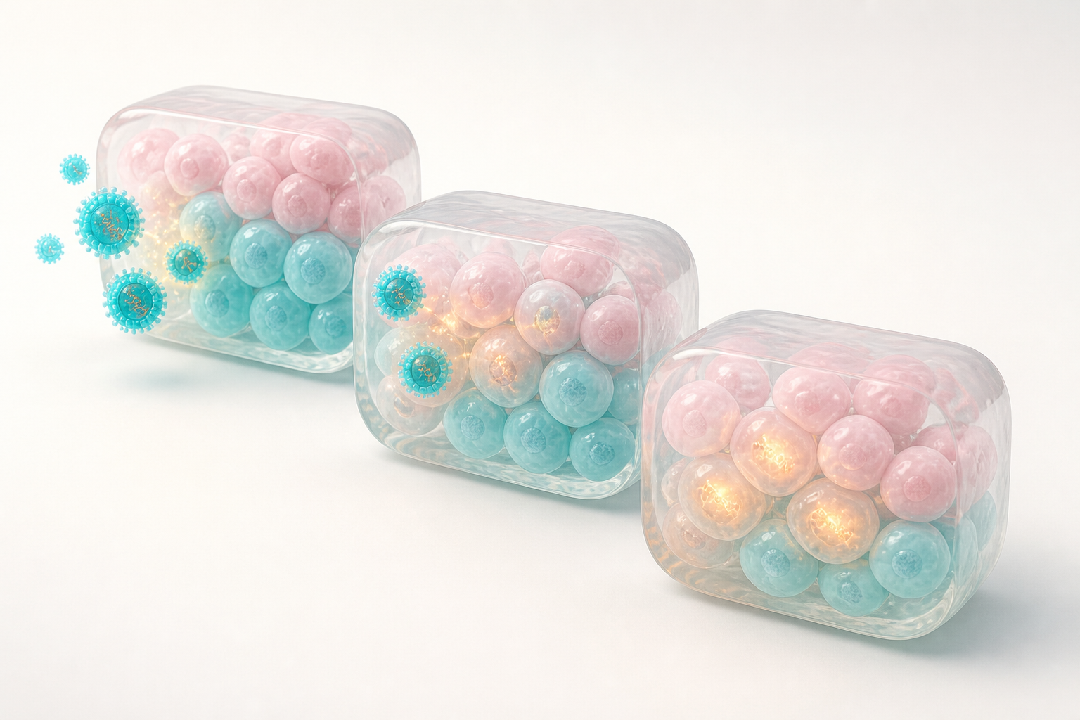
A single siRNA animation can solve one communication problem, but a reusable visual system can serve a platform. The same delivery vehicle, cell material, RISC complex, RNA colors and knockdown language can be reused across target programs. That consistency helps audiences recognize what is platform-wide and what is program-specific.
Reusable assets also reduce production friction. A team can create a core delivery scene, a RISC loading scene and a knockdown evidence scene, then adapt target labels, tissue context and biomarker output for each program. This is especially valuable for companies with multiple liver, CNS, ocular, oncology or rare disease candidates that share delivery logic but differ in target biology.
Commercially, this keeps scientific communication coherent across investor decks, BD packages, website visuals and conference material. It also helps internal teams avoid rebuilding the same mechanism from scratch. The most useful system is flexible enough for new indications while still recognizable as the company's platform language.
- Define which colors represent carrier, guide strand, passenger strand and target mRNA.
- Keep delivery, RISC and knockdown scenes modular for reuse.
- Create still renders from animation scenes for decks and web pages.
Storyboard for siRNA MOA Figures and Animation
A storyboard prevents a siRNA therapeutic mechanism of action animation from becoming a collection of attractive shots. Each scene should have a biological job, a viewer takeaway and a clear handoff to the next step. This also makes review easier because scientists can approve claims scene by scene.
The table below is a practical starting point for biotech teams planning siRNA MOA figures, 3D renders or a short mechanism animation.
| Scene | Visual | Motion | Scientific purpose |
|---|---|---|---|
| Delivery context | Target tissue with a distinct carrier object | Carrier approaches selected cells | Show how the therapeutic reaches the relevant cell population |
| Cellular uptake | Translucent membrane and intracellular compartment | Carrier enters through a controlled internalization beat | Connect delivery vehicle to productive intracellular access |
| Cargo release | siRNA duplex exits the compartment | Warm signal glows guide the release path | Explain how cargo becomes available for RNA interference |
| RISC loading | Simplified protein complex holding guide strand | Passenger strand separates as the guide remains | Make sequence-guided silencing understandable |
| Target knockdown | Complementary mRNA aligns, cleaves and fades | Protein output or biomarker signal decreases | Link molecular mechanism to evidence and therapeutic rationale |
FAQ About siRNA Therapeutic Mechanism of Action Animation
What should a siRNA MOA animation include?
AA useful animation should include delivery, cellular uptake, endosomal release, RISC loading, target mRNA recognition, mRNA cleavage and the resulting knockdown readout. The level of detail should match the audience and the claim being made.
How long should a siRNA mechanism animation be?
AMany biotech use cases work well as a 45 to 90 second sequence plus still renders. Investor and partner materials often need a shorter version that can be understood without narration, while scientific conferences may support a longer annotated cut.
Can the same siRNA visuals support figures and videos?
AYes. A well-planned 3D scene can produce cover images, section figures, deck renders, website stills and animated shots. Planning the asset system early makes the visual language more consistent and efficient.
How detailed should RISC be?
ARISC should be recognizable as the functional silencing complex, but it does not need atomic detail unless the story depends on structure. In many commercial visuals, simplified geometry is clearer and less likely to imply unsupported precision.
CTA: Plan siRNA MOA Visuals With Animiotics
Animiotics helps biotech, platform and research teams turn siRNA biology into clear scientific figures, polished 3D renders and mechanism of action animation assets. The best starting point is a focused visual plan: delivery context, intracellular mechanism, knockdown evidence and reusable platform scenes.
If your team is preparing an investor deck, partner presentation, launch page, manuscript package or conference visual system, Animiotics can help translate the mechanism into a coherent asset set that looks polished without losing scientific credibility.
Open this template in Animiotics
- Use this workflow when siRNA delivery, RISC loading or knockdown evidence needs to be explained clearly.
- Bring target biology, delivery assumptions and key proof points into the first visual planning pass.