Why Membrane Protein Visualization Services Matter
Membrane protein visualization services help biotech and pharma teams explain targets that are scientifically important but visually hard to read. Receptors, channels, transporters and membrane enzymes sit inside a lipid environment, move between states and interact with ligands, antibodies, ions, lipids or intracellular partners. A raw structure screenshot rarely explains why that biology matters to a buyer.
The commercial challenge is simple. A platform team may have strong membrane protein data, but the audience still needs to see the target, the membrane context, the binding event and the mechanism consequence. If those pieces stay abstract, a strong discovery story can sound like another technical claim rather than a credible program advantage.
Animiotics builds premium 3D scientific renders and animations for teams that need membrane protein mechanisms to work on websites, investor decks, pharma partnering materials, conference loops and research presentations. The goal is not decorative science art. The goal is clear visual evidence that helps complex target biology become easier to evaluate.
- Show the membrane context before explaining the mechanism.
- Make receptors, channels and transporters readable at a glance.
- Connect target biology to the platform or program decision.
Start With The Membrane Protein Claim
A useful visual starts with one claim. Is the protein a druggable receptor, a gated channel, a transporter with a directional cycle, a membrane enzyme, a viral fusion protein or an antibody target? That choice determines the camera path, the molecular detail and the amount of supporting context the audience needs.
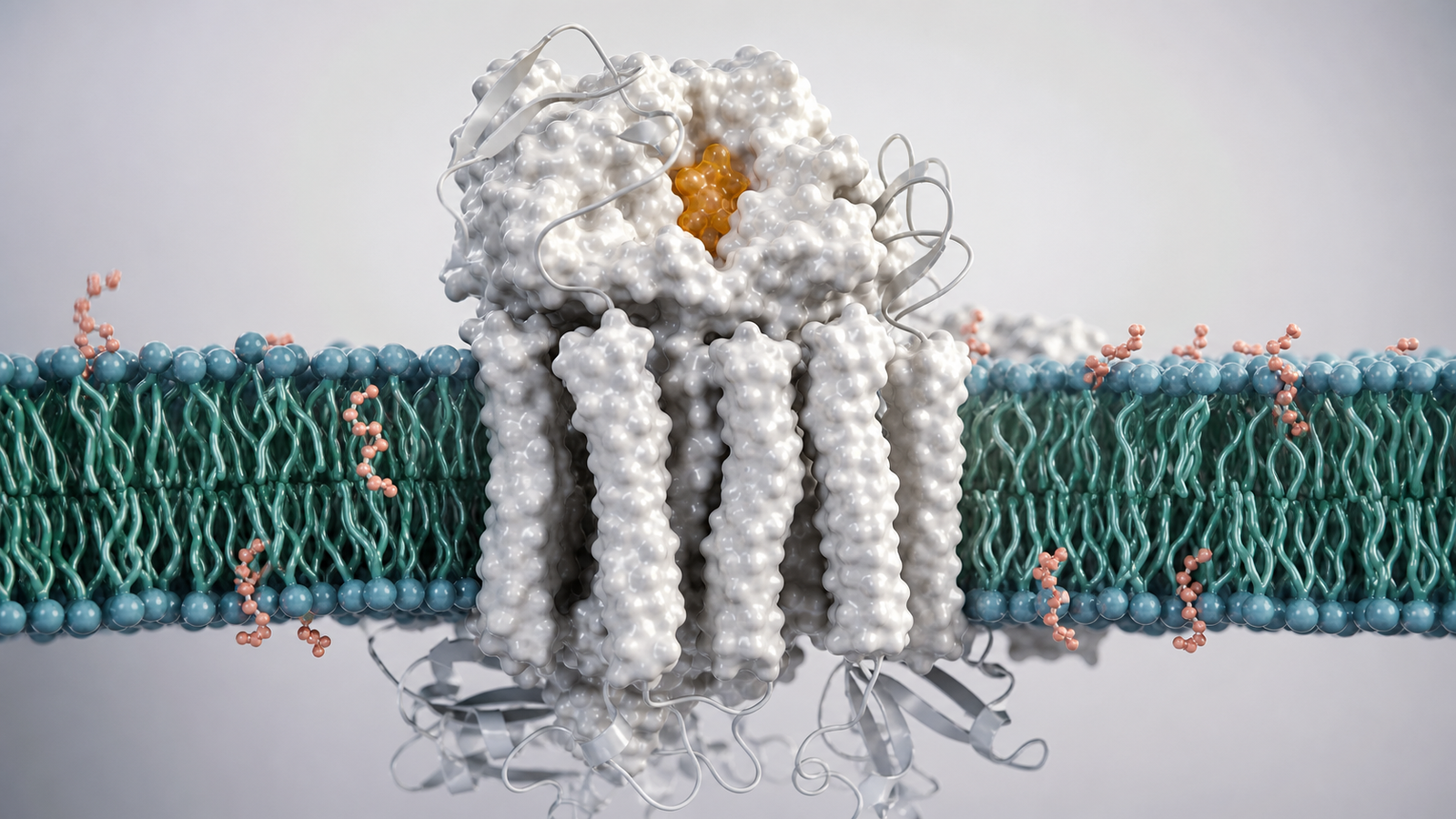
Many teams open with a complete molecular assembly because the structure is impressive. That can help a specialist audience, but buyer-facing communication usually needs a cleaner sequence. A wide hero render can establish the lipid bilayer, then isolate the membrane protein surface, then focus on the ligand, ion pore, epitope or conformational state that carries the commercial story.
This is where membrane protein visualization services become more than rendering support. The visual system can turn a difficult target into a guided explanation. A receptor can shift from inactive to active. A channel can open around a readable pore. A transporter can show substrate movement across the membrane. A binding pocket can become the proof point rather than a hidden detail.
- Define the target claim before building the scene.
- Use the membrane as scientific context, not background decoration.
- Give the viewer one molecular event to remember first.
What Buyer-Ready Membrane Protein Visuals Should Show
Buyer-ready membrane protein visuals usually need four layers. The first layer is the target in its environment: protein, lipid bilayer, extracellular region and intracellular region. The second layer is the mechanism: ligand binding, ion conductance, substrate transport, signaling, inhibition or activation. The third layer is the evidence: structure, cryo-EM density, binding data, assay response or model confidence. The fourth layer is the business consequence: why this target, molecule or platform is worth attention.
Those layers should be sequenced instead of crowded into one frame. A polished animation might start with a membrane surface, reveal a receptor as the hero subject, show a ligand entering the pocket, then move to the intracellular response. A transporter story might show substrate recognition, occlusion and release across the bilayer. A channel story might use a simple ion stream to make gating obvious without adding labels.
For related context, see https://animiotics.com/blog/cryo-em-visualization-services-how-to-explain-structures-conformations-and-binding-evidence-clearly/ and https://animiotics.com/blog/structure-based-drug-design-visualization-services-how-to-explain-pockets-poses-sar-and-platform-value-clearly/. Those posts focus on structural evidence and binding pockets, while membrane protein visualization focuses on making the target readable inside the biological environment where the mechanism happens.
A reusable visual system matters because membrane protein stories often need several formats. The same target model can support a website hero, a mechanism animation, a deck still, a conference loop, a social crop and a partner-specific target variant. Planning for those uses early keeps the visuals consistent while giving the team more commercial value from the same scientific asset.
- Show environment, mechanism, evidence and decision value in order.
- Separate the target from surrounding lipids with restrained material contrast.
- Use one clean molecular event per scene.
Visualizing Receptors Channels And Transporters
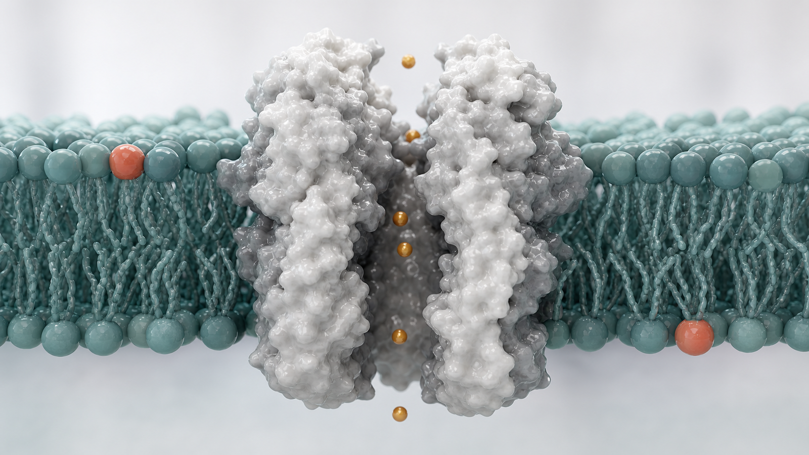
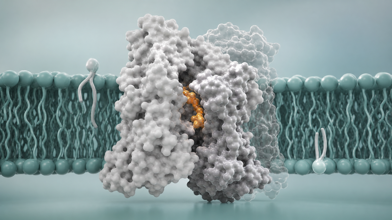
Different membrane proteins need different visual grammar. Receptors often need ligand binding, dimerization, conformational shift or downstream signaling. Channels need a readable pore, gating state and ion movement. Transporters need direction, substrate recognition and alternating access. Membrane enzymes may need an active site, substrate path and lipid context.
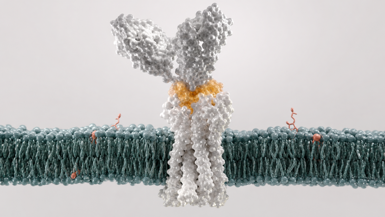
The visual should respect those differences without becoming a technical diagram. A GPCR, kinase receptor or immune receptor can be shown as one elegant molecular surface embedded in a clean bilayer, with sparse supporting proteins only where they clarify signaling. An ion channel can use a central pore and a few amber ions to show conductance. A transporter can show a substrate crossing from one side of the membrane to the other with a simple state transition.
The strongest scenes feel physically plausible. Lipids should not look like candy beads. Protein surfaces should not look like plastic toys. Lighting, depth of field and camera movement should make the structure feel crafted in Blender or Maya while keeping the science disciplined. The audience should understand that the render represents a real target class, not a generic molecular collage.
- Use the protein class to choose the visual sequence.
- Keep supporting forms sparse so the target remains the hero.
- Make motion and direction visible without fake UI or labels.
How Premium 3D Helps Explain Binding Evidence
Binding evidence is often the point where a membrane protein story becomes commercially useful. A small molecule, antibody, peptide, lipid, ion or substrate needs to be visible enough for the audience to understand engagement. The visual should show where the molecule binds, what state it stabilizes and why that interaction supports the claim.
A premium 3D scene can move from the full membrane protein to a focused pocket or epitope without losing spatial context. The membrane remains present but softened. The target surface remains readable. The binding partner carries a controlled accent color. A few nearby helices or loops can frame the interaction so the viewer sees both the molecular detail and the larger mechanism.
This is useful for business development and investor communication because the image does not ask the audience to interpret a specialist figure. It turns the binding claim into a visual event. The buyer can see the ligand settle into a pocket, the antibody recognize an extracellular domain or the substrate pass through a transporter path before the text explains why it matters.
- Use close-ups only after the full membrane context is clear.
- Highlight one binding partner with restrained color.
- Connect binding to state, function or platform value.
Deliverables For Membrane Protein Teams
Most teams need a package of assets rather than one image. A target discovery group may need a website hero render, an animation loop explaining mechanism, high-resolution deck stills, transparent-background protein crops and social launch visuals. A platform company may also need modular versions for several targets that share the same scientific language.
A practical scope often includes model cleanup, membrane scene design, target-class storyboard, one focused binding or gating sequence, three to six still renders and exports for web, deck and event screens. If real structural data is available, the production can use it as the foundation. If the program is earlier, references can guide a representative model while keeping claims honest.
The table below maps common communication needs to useful membrane protein visual deliverables.
| Communication need | Useful deliverable | Why it helps |
|---|---|---|
| Website platform proof | Wide membrane protein hero render | Shows the target class instantly without dense technical copy |
| Pharma partnering deck | Binding pocket, epitope or gating sequence | Connects the target to a clear discovery decision |
| Conference loop | Short receptor or channel animation | Makes mechanism visible from a distance |
| Program-specific update | Target variant stills and reusable crops | Adapts the same visual language to new programs |
Common Mistakes In Membrane Protein Visualization
The first mistake is hiding the protein inside too much membrane detail. Lipids are important, but the viewer should not have to search for the target. A clean bilayer can establish context while the protein remains the visual center.
The second mistake is showing every state at once. Receptors, channels and transporters may have several conformations, but a buyer-facing visual should guide the viewer through them. One state transition is usually stronger than a crowded comparison scene.
The third mistake is replacing evidence with decoration. Floating particles, glowing grids, fake dashboards and unreadable labels can make a serious membrane target feel generic. Premium scientific rendering works best when material quality, lighting and camera hierarchy carry the explanation.
The fourth mistake is forgetting the buyer outcome. A membrane protein visual should not stop at showing a beautiful target. It should help explain why the target is druggable, why the mechanism is differentiated, why the platform can work or why a specific asset deserves attention.
- Do not let the bilayer obscure the target.
- Do not stack too many conformations into one scene.
- Do not use generic biotech effects when the mechanism needs clarity.
FAQ About Membrane Protein Visualization Services
What do membrane protein visualization services include?
AA strong scope can include scientific story planning, model preparation, membrane scene design, storyboard development, premium 3D rendering, animation, review rounds and final exports for websites, decks, publications, conferences and campaigns.
Can visuals use real structures?
AYes. Production can start from PDB structures, cryo-EM models, AlphaFold-style predictions, internal models or approved reference structures. The final visual should match the scientific claim and avoid overstating unknown detail.
How long should a membrane protein animation be?
AA website loop may only need 10 to 20 seconds. A partner-facing explanation often works best at 45 to 90 seconds when it moves from target context to binding, state change and platform value.
What makes membrane protein visuals credible?
ACredible visuals use clear hierarchy, physically plausible biomolecular materials, restrained color, polished lighting and purposeful camera movement. They avoid fake labels, crowded figure panels and toy-like molecular forms when the audience needs a clean commercial story.
Ready To Make Membrane Protein Biology Clear
Membrane protein visualization services are most valuable when they turn a difficult target into a story buyers can follow. The right visual system shows the target in context, makes the mechanism visible, connects binding or state evidence to value and gives the team reusable assets for web, deck and partner communication.
Animiotics creates premium 3D scientific renders, mechanism animations and structural biology visual systems for biotech, pharma, platform and research teams. The work is designed to help receptors, channels, transporters and membrane mechanisms become clear outside the specialist group.
If your membrane protein program, receptor platform, ion channel story or transporter mechanism needs to be clear in a website, deck or partner meeting, start with Animiotics and turn target biology into visuals buyers can understand.
- Explain target context without overwhelming the viewer.
- Turn binding and state changes into readable visual events.
- Build reusable membrane protein visuals for web, deck and partner use.