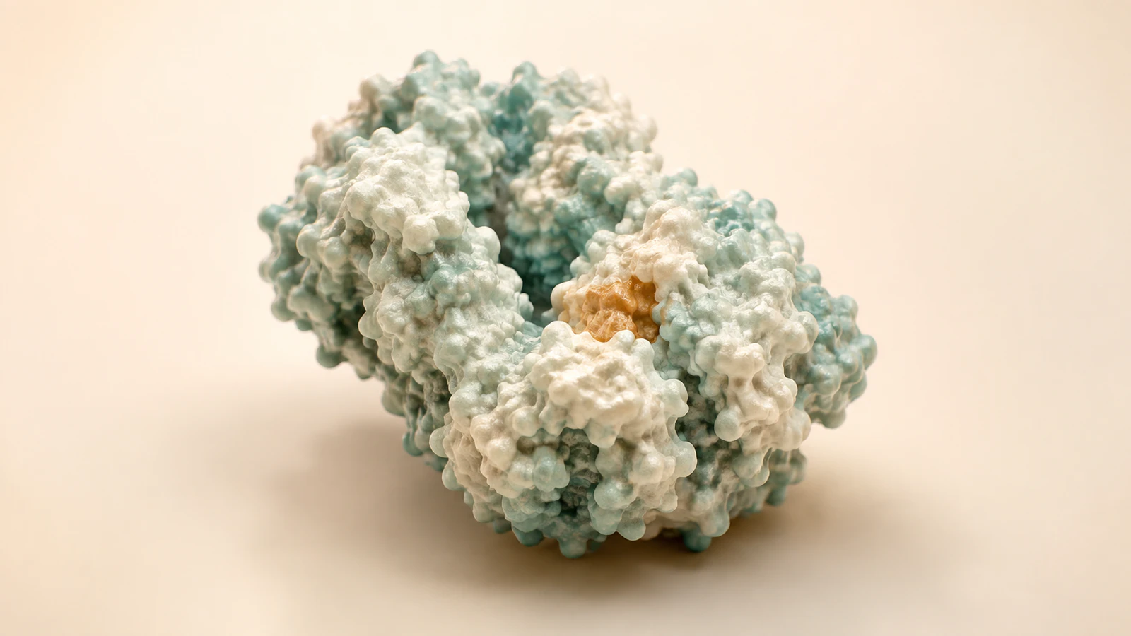
Show a side-chain link, then show what changes
A clear disulfide bond animation follows two cysteine side chains before, during, and after a change in their connection. Keep the protein backbone traceable, identify the sulfur atoms, and make the chemical event visible at a useful camera distance. A glowing bridge alone cannot tell the viewer whether a bond formed, changed partners, or was reduced.
This guide is for scientists and animators preparing a short protein mechanism sequence. Its practical deliverable is a bond ledger paired with a five-shot storyboard. You can use the ledger to review an existing movie or to specify a new one before investing in detailed materials and motion. The central review question is simple: which connection changed, and which connections stayed intact?
The accompanying renders are conceptual illustrations. Their protein shapes, spacing, colors, and camera choices are explanatory, not coordinates from an experiment. Use a documented structure for a named protein, and reserve an explicitly simplified model for teaching the general connection.
Locate the sulfur atoms without losing the backbone
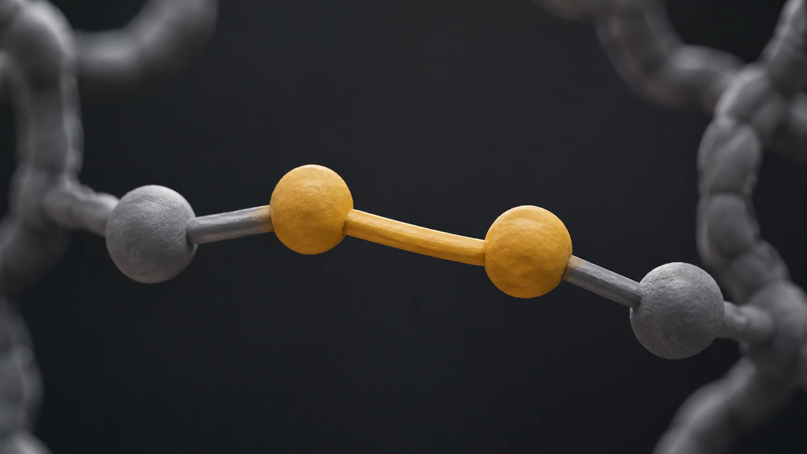
A disulfide is a covalent connection between the sulfur atoms of two cysteine residues, meaning cysteine units within a protein chain. A peptide bond instead joins successive amino-acid residues along that chain. These definitions give the scene two different kinds of continuity to preserve. See the EMBL-EBI protein-structure glossary for the underlying terminology.
Build a local view that lets the eye travel from one backbone position along its side chain, across the sulfur-to-sulfur bond, and back along the second side chain. Avoid placing the sulfur spheres directly into the ribbon path. That shortcut can make the disulfide appear to replace a section of the backbone, especially when the camera hides the branch points.
For a small screen, reduce surrounding detail before enlarging the bond into a long cable. A tight crop and restrained highlight can expose the connection while keeping the local geometry credible. In a deliberately schematic model, state that bond lengths and atom sizes are illustrative.
Decide whether the link is within one chain or between chains
An intrachain link connects two positions within one polypeptide chain; an interchain link connects different chains. Establish that distinction in the opening view, before adding motion. Give each chain a consistent visual identity and keep a reference view available while reviewing the close-up. A cross-link should never create uncertainty about how many backbones are present.
In the bond ledger, identify both partners with chain identifier, residue identifier, and atom name from the chosen source. Record any difference between the numbering used in the paper and the numbering in the structure file. Do not assume that two residues sharing a number in different chains represent the same site.
If the brief says only “show the disulfides,” return to the biological question. A general introduction may need one representative link. A mutation explanation may require one exact residue pair. An assembly story may need multiple chains visible together. Those are different scenes, with different review requirements.
Choose a biological setting for formation or rearrangement
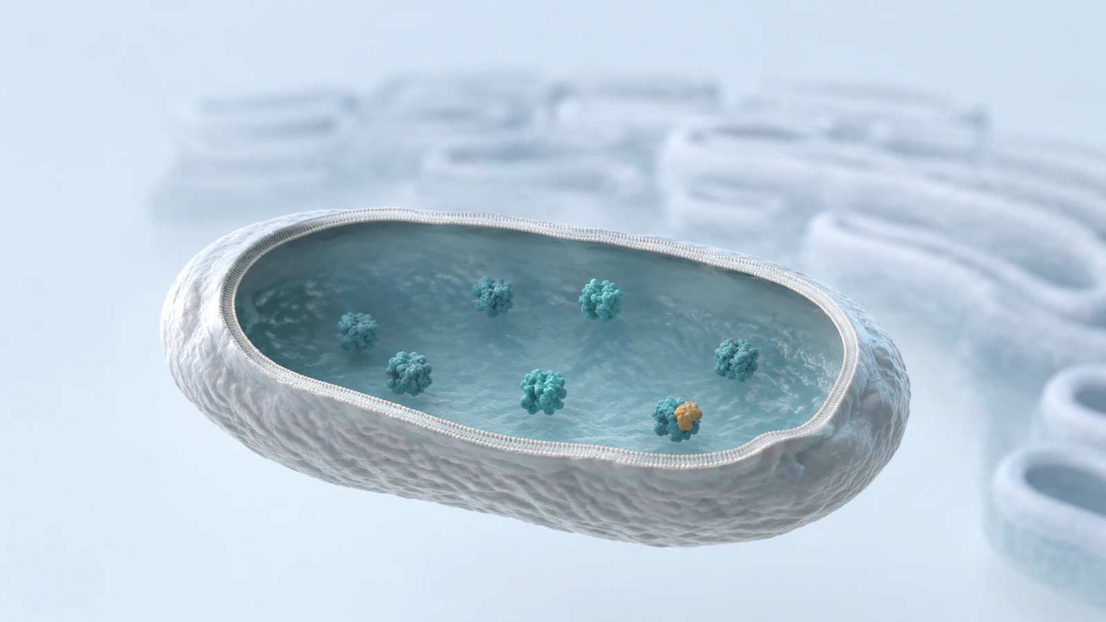
For many eukaryotic proteins entering the secretory pathway, the endoplasmic reticulum, or ER, provides the cellular setting for folding and disulfide formation. The relevant interior compartment is the ER lumen. The cytosol is generally reducing, so it should not be used as an interchangeable backdrop for this story. This context is described in The Cell: The Endoplasmic Reticulum.
A short establishing shot can show a membrane surrounding the lumen, then move to the protein of interest. A cutaway is useful if its removed wall is recognizable as an illustration choice. Keep nearby molecules sparse enough to preserve the compartment boundary. There is no need to invent concentrations, crowding levels, or a measured spatial arrangement.
If the subject is a purified protein undergoing a laboratory treatment, use that experimental context instead. Do not reuse an ER background simply because it looks biological. Record the system in the brief: cellular folding, a defined sample condition, or a general chemistry explanation.
Give rearrangement its own visual grammar
Protein disulfide isomerase, abbreviated PDI, can catalyze the breaking and re-forming of disulfide connections during protein folding. Its role includes helping cysteine partners rearrange toward a pairing compatible with the folded protein. This established mechanism is described in The Cell: Protein Folding and Processing.
For an introductory animation, a transition between two clearly identified pairing states may be sufficient. If you show the catalytic exchange itself, obtain the substrate-specific evidence and review every transient connection. Adding a PDI-shaped surface beside a protein does not establish a docking pose or prove the route between two structures.
Use a separate annotation in the production notes for every interpolated motion. An observed starting state and an observed endpoint do not automatically supply the path between them. Avoid a neat sequence in which every possible pair forms, fails, and tries again unless that sequence is the intended conceptual analogy and is described as such.
Use a bond ledger with keep, change, and unknown fields
Create one row per connection the audience needs to understand. The row should name the starting partners, the intended endpoint, and the evidence supporting the transition. Add three review fields: keep, change, and unknown. This separates the information your animation must preserve from the motion you are choosing for explanation.
In a generic reduction example, keep means the peptide backbone and side-chain attachments remain continuous. Change means the selected sulfur-to-sulfur connection is absent in the reduced endpoint. Unknown might include the exact conformational response under the chosen conditions. Do not fill that unknown field with a dramatic unfolding effect just to create a satisfying ending.
The ledger also makes revisions manageable. If a reviewer changes the residue assignment, update the row and its affected shots. If the biological endpoint changes, revisit the endpoint first, then adjust the transition. Lighting revisions should not silently alter which atoms or chain segments viewers interpret as connected.
| Ledger field | What to record | Review question |
|---|---|---|
| Identity | Source, chain, residue pair, selected bond | Can another scientist locate this exact pair? |
| Keep | Backbone and unaffected attachments | Did any unrelated connection disappear? |
| Change | Formation, partner exchange, or reduction | Does the visible endpoint match the claim? |
| Unknown | Unmeasured motion, timing, or response | Is illustrative motion clearly identified? |
Work through a five-shot reduction storyboard
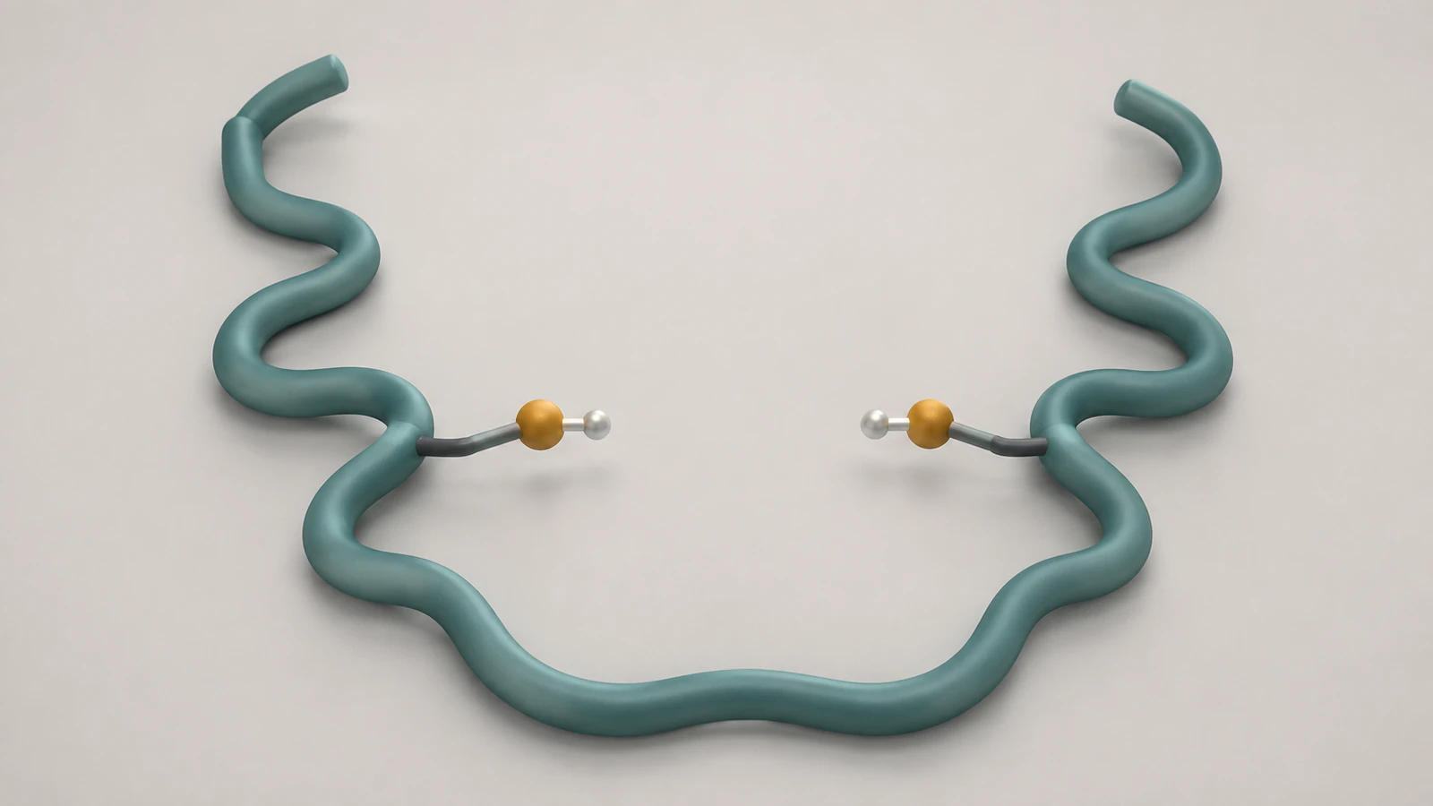
Use a simplified single-chain model for this worked example, not a claim about a particular therapeutic protein. Begin with a visible intrachain cross-link. End with the same continuous backbone and two separated sulfur-containing side groups. The open endpoint drawing is a teaching arrangement, not a prediction that reduction forces every protein into that shape.
Keep the camera position stable during the key connectivity change. If the viewpoint, surface opacity, and bond state all change together, a reviewer cannot tell whether a connection was removed or merely hidden. Make one explanatory change at a time and hold the endpoint long enough to inspect it.
The shot durations belong to your communication plan. Unless a source supplies a relevant measured timescale, do not turn the edit timing into an implied reaction rate. Review the sequence once with narration and once silently; the second pass reveals whether geometry carries the intended distinction.
- Shot 1: Establish the whole chain and the region containing the selected cross-link. Keep chain identity visible.
- Shot 2: Move to the two cysteine side branches. Show where both branches leave the uninterrupted backbone.
- Shot 3: Isolate the sulfur-to-sulfur connection with a restrained color accent. Explain the selected reduction context in narration.
- Shot 4: Show the reduced endpoint with that cross-link absent. Preserve side-chain attachments and backbone continuity; omit unsupported intermediate chemistry.
- Shot 5: Pull back to the same orientation used at the start. Let the viewer compare connectivity without inferring an unmeasured activity change.
Check a real structure before borrowing its identity
A familiar molecule can help reviewers orient themselves, but its identity must remain accurate. For example, RCSB PDB entry 4INS describes pig insulin determined by X-ray diffraction at 1.50 angstrom resolution. It lists A-chain and B-chain entities of 21 and 30 residues, respectively, and several assembly choices. It is not automatically the correct asset for a human insulin product story.
For a named structure, record the entry version, selected assembly, chain identifiers, and the actual disulfide annotations you use. Confirm whether the displayed file contains one molecular unit or a larger assembly. Then inspect the selected residues in a molecular viewer rather than inferring bond identity from a thumbnail.
The PDB-to-animation guide covers the broader structure preparation workflow. Here, the extra requirement is a traceable list of cross-links. Neither a crystal structure nor this article’s generated imagery provides a measured reduction trajectory.
Review representation, chemistry, and interpretation separately
First inspect representation: can the viewer see the branch points and follow each backbone? A molecular surface can establish the overall protein shape but may hide the bond entirely. A local atom view can reveal the chemistry but lose chain context. The surface-versus-ribbon comparison explains how to choose between these views for the question at hand.
Next inspect chemistry against the ledger. Check every endpoint, not only the prettiest middle frame. Look for a sulfur atom detached from its side chain, a cross-link joining the wrong partners, or a backbone segment disappearing under an opacity transition. These are substantive errors even when the final render looks polished.
Finally inspect interpretation. A bond highlight should not imply greater activity, a surface color should not imply a measured redox potential, and a conformational change should not imply a demonstrated functional outcome. Keep any such claims attached to their own evidence rather than letting the visual treatment supply them.
FAQ: disulfide bond animation
Should disulfide reduction cut the protein ribbon?
No. In this teaching sequence, the ribbon represents the peptide backbone, so it remains continuous while the selected side-chain cross-link changes. Backbone cleavage would communicate a different chemical event.
Can I use one gold bridge for every protein?
Use it only as a clearly identified conceptual symbol. For a named protein, verify the cysteine partners and their structural context, then show the selected connection in a representation that preserves those identities.
Does a PDI model prove a folding pathway?
No. A model can locate the enzyme within an explanation, but a specific interaction pose or sequence of intermediates needs its own evidence. Mark transitions that are illustrative.
Should the reduced protein always unfold in the animation?
Do not make that the default ending. Show only the structural response supported for your system, or stop at a clear bond-state comparison that does not claim a particular conformational outcome.
Are the images here exact molecular structures?
No. They are conceptual scientific CGI frames. The local sulfur illustration teaches connectivity, while the protein and ER images provide context; none is a coordinate-derived structure or experimental trajectory.
Try a focused protein animation in Animiotics
Start with one continuous protein chain, one selected cross-link, and one clearly defined endpoint. Use the five-shot plan to explain how the visible connection changes while the backbone remains intact. Bring your bond ledger into review so scientific decisions stay attached to the sequence as the presentation develops.
Explore Animiotics from the homepage and use this focused scene as the starting point for your next scientific animation.